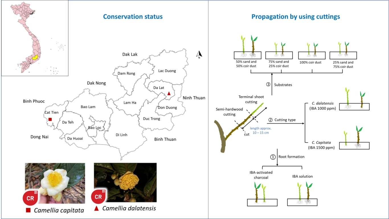
MERCURY CHLORIDE (HgCl2) EXPOSURE CHANGES THE HISTOPATHOLOGICAL FIGURE OF EYE AND BRAIN OF TILAPIA FISH (Oreochromis mossambicus)

Downloads
Mercury pollution brings harmful effects to aquatic animals, the environment and eventually to human health. Mercury accumulates in the liver, kidney, eye lens and brain of fish, resulting in organ damage. This study aimed to determine the effect of HgCl2 exposure on anatomical pathology and histopathology of tilapia fish eye and brain. A total of 36 male tilapia fish were allotted into 4 treatment groups with 3 replications. Fish were exposed to 0.00, 0.25, 0.50, and 0.75 ppm of HgCl2 for 10, 20, and 30 days. Subsequently, the anatomical pathology was observed followed by histopathological examination. Anatomical pathology examination of fish eye on day 30 showed white membrane on the eye lens surface, pupil diminution, and sunken eyes. The brain demonstrated hemorrhage, necrosis, discolorations, and granulated area. The retina showed necrosis, retina pigmentation flexiform layer widened, and cone cell atrophy. Brain depicted structural and cellular damage such as degeneration necrosis and vacuolation. HgCl2 exposure changes the anatomical pathology and histopathology of tilapia fish eye and brain.
Downloads

This work is licensed under a Creative Commons Attribution-NonCommercial-NoDerivatives 4.0 International License.
Authors who publish with this journal agree with the following terms:
- Authors retain copyright and grant the journal right of first publication, with the work 1 year after publication simultaneously licensed under a Creative Commons attribution-noncommerical-noderivates 4.0 International License that allows others to share, copy and redistribute the work in any medium or format, but only where the use is for non-commercial purposes and an acknowledgement of the work's authorship and initial publication in this journal is mentioned.
- Authors are able to enter into separate, additional contractual arrangements for the non-exclusive distribution of the journal's published version of the work (e.g., post it to an institutional repository or publish it in a book), with an acknowledgement of its initial publication in this journal.
- Authors are permitted and encouraged to post their work online (e.g., in institutional repositories or on their website) prior to and during the submission process, as it can lead to productive exchanges, as well as earlier and greater citation of published work (See The Effect of Open Access).