HEPATOPROTECTIVE EFFECT OF Physalis angulata L. LEAF ETHANOL EXTRACT AGAINST BUSULFAN-INDUCED LIVER DAMAGE IN FEMALE RATS

Downloads
Additional Files
ARTICLE HIGLIGHTS
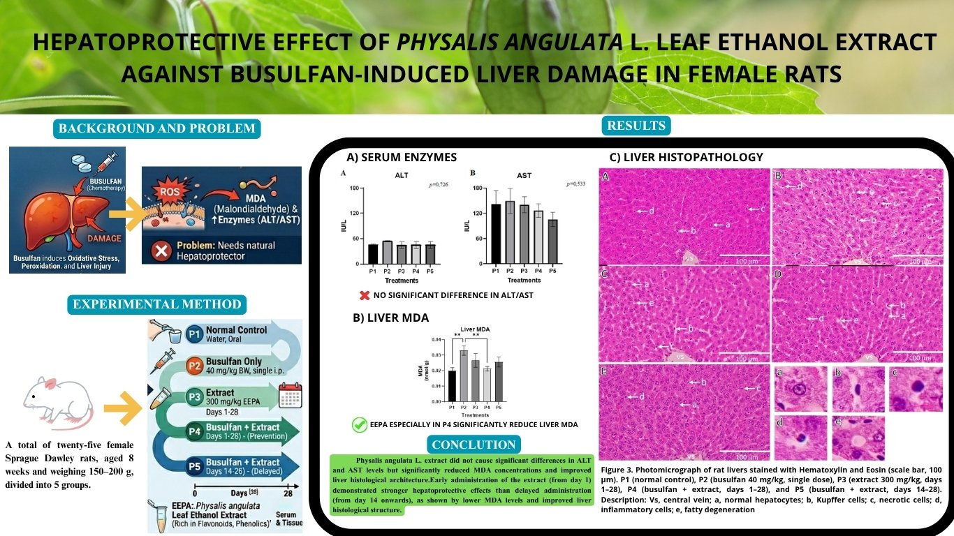
Busulfan can damage the liver by triggering oxidative stress
Physalis angulata leaves show natural liver protective effects
Extract reduced oxidative damage in liver tissue
Liver structure improved after extract treatment
Early treatment provided stronger liver protection than delayed use
ABSTRACT
Liver disease is the leading cause of death worldwide, with drug-induced hepatotoxicity representing a major contributing factor. Busulfan, an anticancer drug used in chronic myeloid leukemia, is cytotoxic to proliferating cells and can induce liver injury. Physalis angulata L. (ground cherry) contains bioactive compounds with potential hepatoprotective properties. This study aimed to evaluate the protective effects of ethanol extract of Phyasalis angulata L. (EEPA) leaves on busulfan-induced liver damage in female Sprague–Dawley rats. Twenty-five rats were randomly divided into five groups: P1 (normal control), P2 (busulfan 40 mg/kg BW, single dose), P3 (extract 300 mg/kg BW, days 1–28), P4 (busulfan + extract, days 1–28), and P5 (busulfan + extract, days 14–28). Parameters assessed were alanine aminotransferase (ALT) and aspartate aminotransferase (AST), the level of malondialdehyde (MDA) in liver, and liver histology. Data were analyzed using one-way analysis of variance (ANOVA) followed by a post hoc test. The results showed that Physalis angulata L. extract did not cause significant differences in the serum ALT and AST levels but significantly reduced the MDA concentrations and improved the liver histological architecture. Busulfan administration significantly decreased hepatocyte count and increased Kupffer and inflammatory cells, fatty degeneration, and necrosis. Early administration of the extract (P4) demonstrated stronger hepatoprotective effects compared with delayed administration (P5). These findings suggest that EEPA leaves exerts hepatoprotective effects against busulfan-induced liver injury in rats, likely through antioxidant activity, as evidenced by subsequent histological improvement, and highlighting its potential as a natural hepatoprotective agent.
Alfarisi H, Wresdiyati T, Sadiah S, Juliandi B. 2022. Nanoextract of Acalypha hispida leaves increases antioxidant defense and suppresses microstructure damage in liver and kidney of diabetic rats. J Appl Pharm Sci 12(10):99–108. DOI:10.7324/JAPS.2022.121010 DOI: https://doi.org/10.7324/JAPS.2022.121010
Angela BT, Astuti DR, Adnyane IKM, Satyaningtijas AS. 2023. Ciplukan leaf extract (Physalis angulata L.) can potentially improve kidney function. Acta Vet Indones 11(1):9–16. DOI:10.29244/avi.11.1.9-16 DOI: https://doi.org/10.29244/avi.11.1.9-16
Balakrishnan B, Illangeswaran RSS, Rajamani BM, Arunachalam AK, Pai AA, Mohanan E, …, Balasubramanian P. 2023. Metformin pre-treatment ameliorates busulfan-induced liver endothelial toxicity during hematopoietic stem cell transplantation. PLoS One 18(10):e0293311. DOI: 10.1371/journal.pone.0293311 DOI: https://doi.org/10.1371/journal.pone.0293311
Devarbhavi H, Asrani SK, Arab JP, Nartey YA, Pose E, Kamath PS. 2023. Global burden of liver disease: 2023 update. J Hepatol 79(2):516–537. DOI:10.1016/j.jhep.2023.03.017 DOI: https://doi.org/10.1016/j.jhep.2023.03.017
Ding N, Lu Y, Cui H, Ma Q, Qiu D, Wei X, …, Cao N. 2020. Physalin D inhibits RANKL-induced osteoclastogenesis and bone loss by regulating calcium signaling. BMB Rep 53(3):154–159. DOI:10.5483/BMBREP.2020.53.3.147 DOI: https://doi.org/10.5483/BMBRep.2020.53.3.147
Gęgotek A, Skrzydlewska E. 2019. Biological effects of protein modification by lipid peroxidation products. Chem Phys Lipids 221(2):46–52. DOI:10.1016/j.chemphyslip.2019.03.011 DOI: https://doi.org/10.1016/j.chemphyslip.2019.03.011
Hao Y, Fang H, Yan X, Shen W, Liu J, Han P, …, Feng Y. 2023. Alginate oligosaccharides repair liver injury by improving anti-inflammatory capacity in a busulfan-induced mouse model. Int J Mol Sci 24(4). DOI:10.3390/ijms24043097 DOI: https://doi.org/10.3390/ijms24043097
Kalas MA, Chavez L, Leon M, Taweesedt PT, Surani S. 2021. Abnormal liver enzymes: A review by clinicians. World J Hepatol 13(11):1688–1698. DOI:10.4254/wjh.v13.i11.1688 DOI: https://doi.org/10.4254/wjh.v13.i11.1688
Komolkriengkrai M, Nopparat J, Vongvatcharanon U, Anupunpisit V, Khimmaktong W. 2019. Effects of glabridin on collagen deposition in the liver and amelioration of hepatocyte destruction in diabetic rats. Exp Ther Med 17(6):1164–1174. DOI:10.3892/etm.2019.7664 DOI: https://doi.org/10.3892/etm.2019.7664
Ma X, Yuan J, Liu X, Xu J, Han J, Wang X, Zhao L. 2023. Busulfan-induced hepatic sinusoidal endothelial cell injury: The modulatory role of pirfenidone for therapeutic purposes. Toxicol In Vitro 92(10):105663. DOI: 10.1016/j.tiv.2023.105663 DOI: https://doi.org/10.1016/j.tiv.2023.105663
Mas-Bargues C, Escrivá C, Dromant M, Borrás C, Viña J. 2021. Lipid peroxidation was measured by chromatographic determination of malondialdehyde. Human plasma reference values for health and disease. Arch Biochem Biophys 709. DOI:10.1016/j.abb.2021.108941 DOI: https://doi.org/10.1016/j.abb.2021.108941
Meira CS, Soares JWC, dos Reis BPZC, Pacheco LV, Santos IP, Silva DKC, …, Soares MBP. 2022. Therapeutic applications of physalins: Powerful natural weapons. Front Pharmacol 13(4):1–14. DOI:10.3389/fphar.2022.864714 DOI: https://doi.org/10.3389/fphar.2022.864714
Melchart D, Hager S, Albrecht S, Dai J, Weidenhammer W, Teschke R. 2017. Herbal traditional chinese medicine and suspected liver injury: A prospective study. World J Hepatol 9(29):1141–1157. DOI:10.4254/wjh.v9.i29.1141 DOI: https://doi.org/10.4254/wjh.v9.i29.1141
Moghadam MT, Dadfar R, Khorsandi L. 2021. Effects of ozone and melatonin on busulfan-induced testicular damage in mice. J Bras Reprod Assist 25(2):176–184. DOI:10.5935/1518-0557.20200081 DOI: https://doi.org/10.5935/1518-0557.20200081
Qin S, Wang J, Yuan H, He J, Deng Y. 2023. Liver function indicators and risk of hepatocellular carcinoma: A bidirectional Mendelian randomization study. Front Genet 14(1):1–7. DOI:10.3389/fgene.2023.1260352 DOI: https://doi.org/10.3389/fgene.2023.1260352
Qin Y, Liu L, He Y, Wang C, Liang M, Chen X, …, Wang D. 2016. Testicular busulfan injection in mice to prepare recipients for spermatogonial stem cell transplantation is safe and nontoxic. PLoS One 11(2):1–15. DOI:10.1371/journal.pone.0148388 DOI: https://doi.org/10.1371/journal.pone.0148388
Silva-Santana G, Bax JC, Fernandes DCS, Bacellar DTL, Hooper C, Dias AASO, …, Mattos-Guaraldi AL. 2020. Clinical hematological and biochemical parameters in Swiss, BALB/c, C57BL/6, and B6D2F1 Mus musculus mice. Anim Model Exp Med 3(4):304–315. DOI:10.1002/ame2.12139 DOI: https://doi.org/10.1002/ame2.12139
Singh S, Osna NA, Kharbanda KK. 2017. Treatment options for alcoholic and nonalcoholic fatty liver disease: A review. World J Gastroenterol 23(36):6549–6570. DOI:10.3748/wjg.v23.i36.6549 DOI: https://doi.org/10.3748/wjg.v23.i36.6549
Teja PTHS, Arjana AAG, Setiasih NLE, Merdana IM. 2021. the impact of oral administration of minyak rajas on liver histopathology and aminotranspherase activities in male kampong chicken. Indones Med Veterinus 10(2):233–244. DOI:10.19087/imv.2021.10.2.233 DOI: https://doi.org/10.19087/imv.2021.10.2.233
van der Velden WJFM, de Weerd-de Jong EC, de Haan AFJ, Blijlevens NMA. 2018. Busulfan-dependent hepatotoxicity of anti-thymocyte globulin formulations during conditioning for hematopoietic stem cell transplantation. Biol Blood Marrow Transplant 24(2):410–412. DOI:10.1016/j.bbmt.2017.10.020 DOI: https://doi.org/10.1016/j.bbmt.2017.10.020
Wang L, Wu H, Yang F, Dong W. 2019. Protective effects of myricetin against cardiovascular diseases. J Nutr Sci Vitaminol 65(6):470–476. DOI:10.3177/jnsv.65.470 DOI: https://doi.org/10.3177/jnsv.65.470
Wiraswati HL, Ekawardhani S, Rohmawaty E, Laelalugina A, Zuhrotun A, Hendriani R, …, Dewi S. 2024. Antioxidant, anti-inflammatory, and antifibrotic activities of ciplukan (Physalis angulata L) extract. J Inflamm research 17:6297–6306. DOI: 10.2147/JIR.S470318 DOI: https://doi.org/10.2147/JIR.S470318
Wu K, Huang R, Wu H, Liu Y, Yang C, Cao S, …, Wu C. 2016. Collagen-binding vascular endothelial growth factor attenuates CCl4-induced liver fibrosis in mice. Mol Med Rep 14(5):4680–4686. DOI:10.3892/mmr.2016.5826 DOI: https://doi.org/10.3892/mmr.2016.5826
Xiang D, Zou J, Zhu X, Chen X, Luo J, Kong L, Zhang H. 2020. Physalin D attenuates hepatic stellate cell activation and liver fibrosis by blocking TGF-β/Smad and YAP signaling. Phytomedicine 78(1):153294. DOI:10.1016/j.phymed.2020.153294 DOI: https://doi.org/10.1016/j.phymed.2020.153294
Zhao Y, Zhao S, Liu S, Ye W, Chen WD. 2025. Kupffer cells, limelight in liver regeneration. Int Immunopharmacol 146(12):113808. DOI:10.1016/j.intimp.2024.113808 DOI: https://doi.org/10.1016/j.intimp.2024.113808
Copyright (c) 2026 Elsi Rahmadhani, Aryani Sismin Satyaningtijas, Damiana Rita Ekastuti, Ronald Tarigan, I Ketut Mudite Adnyane, Kusdiantoro Mohamad, Wahono Esthi Prasetyaningtyas, Yanti Sulistyana

This work is licensed under a Creative Commons Attribution-NonCommercial-NoDerivatives 4.0 International License.
Authors who publish with this journal agree with the following terms:
- Authors retain copyright and grant the journal right of first publication, with the work 1 year after publication simultaneously licensed under a Creative Commons attribution-noncommerical-noderivates 4.0 International License that allows others to share, copy and redistribute the work in any medium or format, but only where the use is for non-commercial purposes and an acknowledgement of the work's authorship and initial publication in this journal is mentioned.
- Authors are able to enter into separate, additional contractual arrangements for the non-exclusive distribution of the journal's published version of the work (e.g., post it to an institutional repository or publish it in a book), with an acknowledgement of its initial publication in this journal.
- Authors are permitted and encouraged to post their work online (e.g., in institutional repositories or on their website) prior to and during the submission process, as it can lead to productive exchanges, as well as earlier and greater citation of published work (See The Effect of Open Access).