Effect of Calcium Nitrate on Chlorophyll-Based Biotransducer Characterization of Arthrospira platensis Gomont

ARTICLE HIGLIGHTS
- Calcium nitrate is utilized to enhance the growth and chlorophyll quality of Arthrospira platensis Gomont, aiming to maximize its potential as a biotransducer molecule.
- Biomass productivity was monitored every three days during a 15-day cultivation period, with specific attention to biomass accumulation and specific growth rates during the stationary phase.
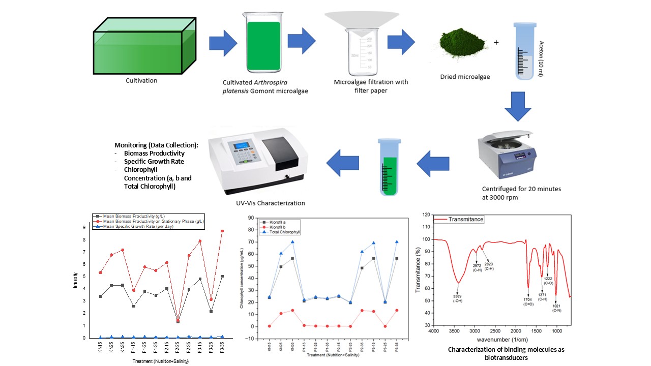
- Chlorophyll concentrations (chlorophyll a, chlorophyll b, and total chlorophyll) were measured using a UV-Vis spectrophotometer at wavelengths of 648 nm and 664 nm.
- Fourier-transform infrared spectroscopy (FTIR) was performed on chlorophyll extracts to assess molecular binding capacity, reinforcing Arthrospira platensis Gomont’s potential as a biotransducer.
- A concentration of 4.5 g/L of calcium nitrate, in combination with 35 ppt salinity, was found to be optimal for enhancing chlorophyll production during cultivation.
ABSTRACT
The aim of this study is to investigate the potential of calcium nitrate as a specific nutrient capable of enhancing the chlorophyll content and optimizating the biotransducer characterization in Arthrospira platensis Gomont. The experimental design employed a Completely Randomized Design (CRD) with Two Factors, consisting of 12 treatments and 3 replications. Each research group was conditioned with varying salinity levels: 15 ppt (S15), 25 ppt (S25), and 35 ppt (S35). In the treatment groups, calcium nitrate was added at different concentrations: 2.5 g/L (P1), 3.5 g/L (P2), and 4.5 g/L (P3). Biomass accumulation and specific growth rate were monitored and data were collected throughout the experiment. At the end of the treatment period, chlorophyll was extracted and its concentration was measured using UV-Vis Spectrophotometry and FTIR analysis. The results indicated that the addition of calcium nitrate 4.5 g/L combined with 35 ppt salinity increased the average biomass productivity over 15 days by 5.1 g/L, with a specific growth rate in the stationary phase of 0.12 per day. Supplementation of calcium nitrate 4.5 g/L in 35 ppt salinity was found to increase total chlorophyll concentration to 70.15 μg/mL, further supporting its potential as a supplementary nutrient to enhance the biotransducer properties with five key functional groups for the stability and binding affinity of analyte molecules in SPR application.
INTRODUCTION
Surface Plasmon Resonance (SPR)-based biosensors have been widely applied in biomedical fields, particularly for the real-time detection of biological analytes. SPR is an optical technique in which changes in the intensity of reflected light at a specific resonance angle on a metal-modified surface are measured. This surface functions as a receptor, with variations in the refractive index near the interface occurring as a result of analyte binding. The analytes may include biomarkers such as DNA, proteins, enzymes, and other biological macromolecules.
In recent years, SPR biosensors have been developed as efficient, cost-effective, and user-friendly alternatives for biomarker detection (Écija-Arenas et al., 2021); (D’Agata et al., 2024). However, conventional SPR systems have continued to face limitations, including suboptimal sensitivity and unstable performance, primarily due to inadequacies in the biointerface sensing layer. These limitations highlight the need for incorporating additional external supporting layers beyond the traditional gold (Au) film and prism to enhance signal transduction and reduce energy loss across the optical interface (SN et al., 2022). Consequently, improvement of the biointerface layer has been identified as a critical priority to advance the performance and reliability of SPR biosensors
In efforts to enhance the performance of SPR biosensors, the potential of chlorophyll as a biotransducer has been identified as an attractive alternative. Chlorophyll, present in large quantities in microalgae such as spirulina (Arthrospira platensisGomont), possesses notable optical properties and molecular interaction capabilities, along with high tolerance and stability under varying environmental conditions, which can improve the sensitivity and specificity of detection systems (Mandal & Dutta, 2020). When incorporated as the primary component of the biointerface sensing layer, chlorophyll is expected to enhance the performance of SPR biosensors in detecting relevant parameters.
Moreover, the selection of calcium nitrate as an additional nutrient during the cultivation phase of Arthrospira platensis Gomont is considered a crucial factor in enhancing chlorophyll production for use as a biotransducer molecule. Calcium nitrate has been reported to influence the growth and metabolism of A. platensis, thereby contributing to increased chlorophyll production (Fakhri et al., 2020). However, the effects of nitrate have been shown to vary among species. (Rani & Maróti, 2021) reported that increasing nitrate concentrations reduced pigment content in Chlamydomonas and Chlorella strains.
In contrast, Chlorella vulgaris achieved higher biomass at elevated nitrate levels, with improved uptake rates of up to 1,798 mg/L and tolerance exceeding 6,014 mg/L (Jeanfils et al., 1993). Nitrate supplementation has also been observed to improve microalgal growth, with biomass concentrations reaching up to 3,188 mg/L (Unknown Author, 2022).
Existing research has largely focused on general microalgal growth, with limited attention given to the targeted enhancement of chlorophyll-based biotransducer properties for biosensing applications. In this study, this gap was addressed through an integrated approach combining physiological optimization under controlled environmental conditions with evaluation of biosensor applicability. Using Arthrospira platensis Gomont as a model organism, the effects of nutrient and salinity modulation on the performance of chlorophyll-based SPR biosensors for metabolic biomarker detection were investigated.
MATERIALS AND METHODS
Material
The initial Arthrospira platensis Gomont inoculum was obtained from a pure culture cultivated by commercial farmers in Tangerang City, Jakarta, Indonesia.
Research Design
A Completely Randomized Design (CRD) with two factors was employed, comprising 12 treatments with 3 replications, resulting in a total of 36 culture groups of Arthrospira platensis Gomont. These groups included a negative control and three treatment sets, each subjected to different salinity levels: 15 ppt (S15), 25 ppt (S25), and 35 ppt (S35).
The negative control was maintained without additional nutrients, whereas the treatment groups were supplemented with calcium nitrate at concentrations of 2.5 g/L (P1), 3.5 g/L (P2), and 4.5 g/L (P3) under the same salinity conditions. Calcium nitrate concentrations were adapted from (Fakhri et al., 2020) with modifications. The experiment was conducted over a 15-day period, with cultures maintained in aquariums under 4,000 lux lighting (24:0 light cycle) and continuous aeration to ensure uniform nutrient distribution and to prevent sedimentation (Fakhri et al., 2020).
Preparation of Culture Media
The culture medium was prepared using sterilized distilled water, treated with 1 mL/L chlorine for 24 hours, and subsequently treated with 1 mL/L sodium thiosulfate for dechlorination. This process was performed to ensure the removal of chlorine residues prior to use in microalgae cultivation (Fakhri et al., 2020).
Inoculation, Cultivation, and Treatment Group Assignment
Inoculation Phase
During the inoculation phase, the initial Arthrospira platensis Gomont inoculum was expanded to obtain the required quantity for cultivation. A volume of 100 mL of the initial inoculum was added to each liter of culture medium. The inoculum was then incubated for 7 days before initiation of the cultivation phase.
Cultivation Phase and Treatment Group Assignment
Following inoculation, 100 mL of Arthrospira platensis Gomont culture was transferred into 2 L of sterile medium, resulting in a 1 : 20 ratio. Cultures were maintained at salinity levels of 15 ppt (S15), 25 ppt (S25), and 35 ppt (S35). Treatment groups were supplemented with calcium nitrate at concentrations of 2.5 g/L (P1), 3.5 g/L (P2), and 4.5 g/L (P3), while the negative control received no nutrient addition. Treatments were applied for 15 days, during which biomass productivity and specific growth rates were monitored and recorded (Fakhri et al., 2020).
Biomass Productivity Assessment
Observations were conducted over a 15-day period, from day 0 to day 15, with measurements taken every three days. Biomass productivity was determined by collecting a 25 mL microalgae sample. The sample was subsequently filtered and dried in an oven at 105 °C for 2 minutes before being weighed. The results were calculated using the following formula (Fakhri et al., 2020):
Arthrospira platensis Gomont biomass (g/L) = \documentclass{article} \usepackage{amsmath} \begin{document} \displaystyle \frac{\text{(B-A)}}{\text{Sample\ volume}}\text{×1}\text{,}\text{000 L\ of\ media} \end{document}
where:
B = Weight of the dried sample and filter paper after being oven-dried at 105 °C for 2 minutes (g);
A = Weight of the filter paper after being oven-dried at 105 °C for 2 minutes (g);
Sample volume = Volume of the sample solution taken from the cultivation medium (mL).
Biomass Productivity Assessment and Specific Growth Rate during the Stationary Phase
Upon reaching the stationary phase, which is characterized by consistent and stable biomass growth as well as constant medium turbidity, typically occurring between days 10 to 15, biomass productivity and specific growth rate were measured using the following formula (Fakhri et al., 2020):
\documentclass{article} \usepackage{amsmath} \begin{document} \displaystyle \mu = \frac{(\ln(X_2) - (\ln(X_1))}{(t_2 - t_1)} \end{document}
where:
μ = Specific growth rate per unit of time (per day);
X₂ = Biomass measured at the final time point (g/L);
X₁ = Biomass measured at the initial time point (g/L)’
t₂ = Final time (days);
t₁ = Initial time (days).
Chlorophyll Extraction
On day 15, 250 mL of microalgae samples were collected from each group and filtered using Whatman No. 1 filter paper with a pore size of 11 μm. The biomass was air-dried, weighed (~1 g), and dissolved in 10 mL of acetone. The solution was transferred to 10 mL bottles, wrapped in aluminum foil to prevent light exposure, and centrifuged at 3,000 rpm for 20 minutes. The supernatant was collected, placed in cuvettes, and its absorbance was measured at 648 nm and 664 nm using a UV-Vis spectrophotometer (Aryono et al., 2022). The concentration of pigments in the extracts was determined using the following equations (Lichtenthaler & Wellburn, 1983):
Chlorophyll a concentration (μg/mL): Ca = (13.36 × A664) -( 5.19 × A648)
Chlorophyll b concentration (μg/mL): Cb = (27.43×A648) - (8.12×A664)
Total Chlorophyll (μg/mL) = Ca + Cb
FTIR (Fourier Transform Infrared Spectroscopy) Analysis
FTIR analysis was conducted using a Shimadzu IR Prestige 21/FTIR 8400 instrument, which was warmed up for at least 30 minutes to stabilize performance. After powering on the detector, the IR Solution software was launched for data acquisition. Approximately 0.5 mL of the liquid sample, free of water or interfering solvents, was applied onto the ATR crystal plate and evenly distributed across its surface. The ATR assembly was then placed into the sample holder. A background spectrum was first obtained to eliminate external interference and ensure accurate sample measurement. Subsequently, the spectrum of the liquid sample was recorded, capturing absorption peaks characteristic of the sample's molecular structure. The resulting spectrum was analyzed using the IR Solution software to identify key absorption bands.
Data Analysis
Biomass increase, specific growth rate, and chlorophyll concentration were statistically analyzed using two-factor ANOVA in SPSS 16 software, followed by Duncan's multiple range test at a significance level of 5% (P = 0.05).
RESULTS AND DISCUSSION
Biomass Productivity of Arthrospira platensis Gomont during 15-day of the Study
Biomass productivity of Arthrospira platensis Gomont was monitored every three days over a 15-day period under various nutritional and salinity treatments (Figure 1a & Figure 1b). At the onset (Day 0), all experimental groups exhibited similarly low productivity, indicating uniform initial conditions. Treatments supplemented with medium to high concentrations of calcium nitrate (P2 and P3) demonstrated a progressive increase in biomass, reaching a peak on Day 15. This trend suggests that elevated nitrate availability supports cellular growth, consistent with previous findings linking nitrogen availability to increased biomass in Chlorella sp. (Unknown Author, 2022).
Unexpectedly, considerable biomass accumulation was also observed in the control group (KN), which received no additional nutrients, particularly on Days 12 and 15 of the cultivation period. This phenomenon may be attributed to adaptive metabolic strategies, such as efficient utilization of residual nutrients or internal nitrogen recycling mechanisms in nutrient-limited microalgal cultures (Bezerra et al., 2020); (Villaró et al., 2023).
Figure 1.Biomass productivity of Arthrospira platensis Gomont during the 15-day study
Biomass productivity was enhanced under higher salinity conditions (35 ppt) compared to lower salinity treatments (15 and 25 ppt), particularly during the final observation period. The combination of 35 ppt salinity and 4.5 g/L calcium nitrate (P3) yielded the highest mean biomass productivity at 5.1 g/L. These findings are consistent with previous studies demonstrating that moderate salinity stress stimulates osmoregulatory mechanisms in microalgae, promoting the synthesis of compatible solutes and enhancing ionic regulation, which collectively support increased biomass production (Bezerra et al., 2020); (Villaró et al., 2023). Furthermore, calcium ions introduced through calcium nitrate are believed to contribute to membrane stabilization and activation of signaling pathways involved in stress tolerance, thereby further improving growth under high-salinity conditions (White & Broadley, 2003); (Unknown Author, 2024).
Despite the observed trends, two-factor ANOVA indicated that the effects of calcium nitrate supplementation (F = 0.611; P = 0.611), salinity (F = 1.579; P = 0.215), and their interaction (F = 1.191; P = 0.324) on biomass productivity were not statistically significant at the 0.05 level Table 1. The adjusted R² value (0.016) further suggested that variability in biomass productivity was only marginally explained by the tested variables. These findings may have been influenced by intrinsic biological variation or the relatively short duration of the experimental period. It is important to emphasize that the absence of statistical significance does not necessarily negate the biological relevance of the observed trends, particularly in microalgal systems where subtle physiological responses may not be captured by conventional significance thresholds (Quinn & Keough, 2002).
Although calcium nitrate supplementation and elevated salinity did not produce statistically significant differences in biomass productivity, the consistent increase observed in treatments P2 and P3, particularly under high salinity, suggests a potential synergistic effect of nitrate availability and osmotic conditioning in enhancing the growth performance of Arthrospira platensis Gomont. Further investigations involving extended cultivation periods, increased replication, and refined physiological measurements are warranted to elucidate the underlying mechanisms and validate these observed trends.
Biomass Productivity of Arthrospira platensis Gomont during the Stationary Phase
The stationary phase typically reflects a condition in which cellular growth slows due to nutrient limitation, waste accumulation, or other environmental constraints (Richmond, 2004). Despite these limitations, sustained or even enhanced biomass productivity was observed in Arthrospira platensis Gomont during the stationary phase (days 10 - 15), particularly under specific combinations of calcium nitrate supplementation and salinity levels (Figure 2 a & b).
Arthrospira platensis Gomont exhibited a substantial increase in biomass productivity, reaching up to 10.8 g/L with an average productivity of 8.8 g/L, particularly in cultures treated with 4.5 g/L calcium nitrate under 35 ppt salinity. This suggests that specific physiological mechanisms supported sustained metabolic activity despite potentially growth-limiting conditions. The enhanced growth is likely attributed to the dual role of calcium nitrate: nitrate (NO₃-) continues to support nitrogen metabolism and chlorophyll synthesis, while calcium (Ca²+) contributes to stabilization of thylakoid membranes, regulation of enzymatic activity, and facilitation of photosynthetic protection mechanisms under stress (Unknown Author, 2024); (The et al., 2021); (Hachiya & Sakakibara, 2017); (Hochmal et al., 2015); (White & Broadley, 2003); (Unknown Author, 2024).
| Source | Type III sum of squares | df | Mean square | F | Sig. |
|---|---|---|---|---|---|
| Corrected model | 79.618a | 11 | 7.238 | 1.103 | 0.375 |
| Intercept | 944.676 | 1 | 944.676 | 143.974 | 0.000 |
| Treatment | 12.021 | 3 | 4.007 | 0.611 | 0.611 |
| Salinity | 20.724 | 2 | 10.362 | 1.579 | 0.215 |
| Treatment*Salinity | 46.872 | 6 | 7.812 | 1.191 | 0.324 |
| Error | 393.687 | 60 | 6.561 | ||
| Total | 1,417.980 | 72 | |||
| Corrected total | 473.304 | 71 |
Figure 2.Biomass productivity of Arthrospira platensis Gomont during the stationary phase
However, this stimulatory effect was not consistent across all treatment groups. Interestingly, the KN group, which received no additional nutrients, achieved comparably high productivity at 35 ppt salinity, highlighting the remarkable adaptive capacity of Arthrospira platensis Gomont. Elevated salinity may act as a mild stressor, triggering metabolic compensation mechanisms that enable continued biomass accumulation despite nutrient limitations (Bezerra et al., 2020); (Villaró et al., 2023).
These findings underscore the interactive effects of nutrient concentration and salinity, where physiological outcomes are nonlinear and dependent on threshold levels and organismal adaptive responses. While high calcium nitrate concentrations combined with elevated salinity promoted growth, intermediate nutrient levels or suboptimal salinity conditions did not elicit the same effect.
In some cases, the absence of supplementation under high salinity conditions paradoxically stimulated biomass accumulation through internal regulatory mechanisms..
Specific Growth Rate of Arthrospira platensis Gomont during the Stationary Phase
The specific growth rate for each treatment group was measured after Arthrospira platensis Gomont entered the stationary phase, as indicated by stable turbidity between Days 10 and 11. This measurement was conducted to evaluate the culture’s capacity for biomass accumulation following the onset of nutritional stress, which results from intensified intra-population competition and depletion of essential nutrients (Richmond, 2004). The specific growth rate results for each group are presented in Figure 3.
Figure 3.Mean specific growth rate of Arthrospira platensis Gomont during the stationary phase
supplemented at concentrations of 3.5 g/L and 4.5 g/L, the specific growth rate increased to 0.11 and 0.12 per day, respectively. The highest growth rate was recorded in the P3-35 group, followed by the P2-35 group. These findings suggest that under elevated salinity, appropriate nitrate supplementation may enhance nitrogen assimilation pathways, support chlorophyll synthesis, and maintain cell division even during the later stages of cultivation (Unknown Author, 2024); (Unknown Author, 2022).
However, this effect was not consistent across all treatment combinations. Groups receiving calcium nitrate at 2.5 g/L (P1), or those cultivated at lower salinities (15 ppt and 25 ppt), exhibited lower specific growth rates. This inconsistency indicates that the physiological response of Arthrospira platensis Gomont to nutrient addition is modulated by salinity, and not all nutrient–salinity combinations yield synergistic effects. Interestingly, the KN-35 group exhibited a growth rate comparable to those of the P2-35 and P3-35 groups. This suggests that Arthrospira platensis Gomont is capable of activating osmoregulatory mechanisms and utilizing internal nutrient reserves to maintain essential metabolic functions despite nutrient constraints, which may contribute to sustaining growth rates under high-salinity stress (Bezerra et al., 2020); (Villaró et al., 2023).
Two-factor ANOVA analysis Table 2 revealed that calcium nitrate supplementation (F = 0.184; P = 0.906), salinity (F = 1.690; P = 0.195), and their interaction (F = 0.416; P = 0.865) did not produce statistically significant differences in specific growth rate (P > 0.05). Although these statistical results do not confirm significant treatment effects, observable patterns suggest that increasing salinity levels (from 15 ppt to 35 ppt) across all treatments tend to correlate with higher biomass productivity and specific growth rates, particularly in the KN and P3 groups. The P2-25 treatment exhibited a significant drop in productivity, which may be attributed to unfavorable salinity-nutrient interactions under this condition. The P3-35 treatment appeared to be the most optimal condition for biomass production, showing the highest values across all metrics, productivity and growth rate, indicating that this specific nutrient-salinity combination may create physiological conditions conducive to enhanced microalgal growth and biomass accumulation.
The data presented in Table 3 suggest that higher salinity levels (35 ppt) generally support enhanced biomass productivity and specific growth rates in Arthrospira platensis Gomont, particularly under the P3 treatment, which consistently outperformed other conditions. However, the interaction between salinity and treatment varied, with the P2-25 condition identified as unfavorable, indicating the necessity for optimization based on specific growth or production targets.
| Source | Type III sum of squares | df | Mean square | F | Sig. |
|---|---|---|---|---|---|
| Corrected model | 0.032a | 11 | 0.003 | 0.585 | 0.832 |
| Intercept | 0.420 | 1 | 0.420 | 84.423 | 0.000 |
| Treatment | 0.003 | 3 | 0.001 | 0.184 | 0.906 |
| Salinity | 0.017 | 2 | 0.008 | 1.690 | 0.195 |
| Treatment*Salinity | 0.012 | 6 | 0.002 | 0.416 | 0.865 |
| Error | 0.239 | 48 | 0.005 | ||
| Total | 0.691 | 60 | |||
| Corrected total | 0.271 | 59 | |||
Note: a: R2 = .118 (Adjusted R2 = .084). |
| Treatment |
Mean biomass productivity (g/L) |
Mean biomass productivity on stationary phase (g/L) |
Mean specific growth rate (per day) |
|---|---|---|---|
| KN15 | 3.4ab | 5.4ab | 0.04a |
| KN25 | 4.3ab | 6.8ab | 0.09ab |
| KN35 | 4.3ab | 7.2ab | 0.10b |
| P1-15 | 2.6ab | 3.9ab | 0.08ab |
| P1-25 | 3.8ab | 5.8ab | 0.10b |
| P1-35 | 3.5ab | 5.5ab | 0.08ab |
| P2-15 | 4.1ab | 6.2ab | 0.06ab |
| P2-25 | 1.3a | 1.5a | 0.06ab |
| P2-35 | 4.0ab | 6.8ab | 0.11b |
| P3-15 | 4.9b | 7.9b | 0.08ab |
| P3-25 | 2.2ab | 3.2ab | 0.09ab |
| P3-35 | 5.1b | 8.8b | 0.12b |
Chlorophyll a content was consistently higher than chlorophyll b across all treatment groups Figure 4. Based on measurements of chlorophyll a and total chlorophyll concentrations, the KN-35, P3-35, P3-15, and P2-35 groups exhibited higher levels compared to other groups. These results indicate that a salinity of 35 ppt combined with calcium nitrate supplementation at concentrations of 2.5 g/L and 3.5 g/L can enhance chlorophyll content.
Figure 4.Chlorophyll concentration in Arthrospira platensis Gomont
When cultivated at 35 ppt salinity without nutrient supplementation, Arthrospira platensisGomont exhibited elevated concentrations of chlorophyll a, chlorophyll b, and total chlorophyll, measured at 56.55, 13.57, and 70.12 μg/mL, respectively. Similarly, under 35 ppt salinity with 4.5 g/L calcium nitrate supplementation, chlorophyll a, b, and total chlorophyll levels were recorded at 56.54, 13.61, and 70.15 μg/mL, respectively. Moreover, in several treatment groups, calcium nitrate supplementation did not result in a significant enhancement of chlorophyll content in Arthrospira platensis Gomont.
Under 15 ppt salinity without nutrient supplementation (KN-15), chlorophyll content in Arthrospira platensis Gomont was relatively low, indicating that suboptimal salinity may not enhance chlorophyll production. Treatments at 25 ppt salinity (KN-25, P1-25, P3-25) showed slight improvements in total chlorophyll compared to KN-15; however, these increases remained lower than those observed in treatments at 35 ppt.
At 35 ppt salinity (KN-35, P2-35, P3-35), increases in chlorophyll content were observed in Arthrospira platensis Gomont. The highest enhancement occurred in the P3-35 treatment, which achieved a total chlorophyll concentration of 70.15 μg/mL. These results suggested that this nutrient-salinity combination provides optimal conditions for biomass growth and chlorophyll accumulation in Arthrospira platensis Gomont.
The results indicated that nutrient supplementation combined with appropriate salinity levels plays a significant role in enhancing chlorophyll content in Arthrospira platensis Gomont. Nitrate, absorbed through NRT1 and NRT2 transporters, supports amino acid andnucleotide biosynthesis essential for cell growth and biomass accumulation. Moreover, nitrate serves as a nitrogen donor for porphyrin ring formation, the core structure of chlorophyll molecules (Unknown Author, 2024), and modulates gene expression under saline stress (The et al., 2021); (Hachiya & Sakakibara, 2017). In calcium nitrate, both nitrate and calcium components contribute to chlorophyll biosynthesis, with calcium stabilizing chloroplast membranes and supporting photosynthetic activity through multiple physiological roles. As a component of the oxygen-evolving complex (Mn₄CaO₅) in photosystem II (PSII), calcium facilitates water splitting and oxygen evolution, while also regulating Calvin cycle enzymes such as fructose-1,6-bisphosphatase (FBPase) and sedoheptulose-1,7-bisphosphatase (SBPase). Under salinity stress, calcium maintains thylakoid membrane integrity and activates protective mechanisms such as cyclic electron flow (CEF) and non-photochemical quenching (NPQ), thereby preserving pigment biosynthesis (Hochmal et al., 2015). These multifaceted roles likely explain the elevated chlorophyll content observed in high-calcium nitrate treatments (notably P3-35), underscoring its potential to enhance photosynthetic efficiency and pigment accumulation under environmental stress
Analysis of Functional Groups in Chlorophyll of Arthrospira platensis Gomont
FTIR analysis revealed that key functional groups essential for biosensor applications were present in chlorophyll extracted from Arthrospira platensis Gomont cultivated with 4.5 g/L calcium nitrate at 35 ppt salinity Figure 5. A strong absorption at 3,389/cm indicated the presence of hydroxyl (–OH) groups, which are known to contribute to hydrogen bonding and enhance molecular interaction stability (Nandiyanto et al., 2023). The carbonyl (C=O) group was detected at 1,704/cm, which is typically found in ester or ketone structures of chlorophyll and is recognized for its role in increasing binding affinity through electrostatic interactions with target molecules (Mansour et al., 2022).
bonds, characteristic of esters or carboxylic acids, which have been found to enhance chlorophyll’s ability to interact with target molecules through polar interactions (LibreTexts, 2024). Meanwhile, the band at 1,021/cm was associated with C–N bonds, which are commonly found in amine or amide groups and the porphyrin structure of chlorophyll. These groups are known to enable electrostatic interactions with charged molecules, thereby strengthening the binding affinity between chlorophyll and target analytes in sensor applications (Mansour et al., 2022).
Overall, these FTIR results indicate that stable chemical interactions with target molecules can be formed by chlorophyll due to the presence of hydroxyl, carbonyl, alkane, C–O, and C–N groups. The role of these functional groups in biosensing applications has been supported by their contribution to enhancing molecular binding affinity. Furthermore, a positive enhancement in chlorophyll production under optimal conditions reinforcing the potential of chlorophyll as a biotransducer for molecular detection (Mansour et al., 2022).
Figure 5.FTIR analysis of chlorophyll from P3-35 group
This study highlights the potential of optimized nutrient conditions to enhance the biotransducer properties of Arthrospira platensis Gomont. Although some treatments did not yield statistically significant results, the increased presence of specific functional groups, as revealed by FTIR analysis, supports their role in strengthening binding affinity in SPR biosensors. Cultivation under appropriate calcium nitrate concentrations and salinity levels was shown to improve chlorophyll content and promote the expression of functional groups favorable for analyte interaction. These findings underscore the potential of Arthrospira platensis Gomont as an effective biotransducer for SPR-based detection systems. Future research should investigate the scalability and real-world applicability of this approach in diagnostic settings. The FTIR-detected functional groups further validate the chemical complexity of chlorophyll and its suitability as a molecular recognition element, particularly due to the presence of groups capable of polar and electrostatic interactions.
CONCLUSION
The addition of calcium nitrate was found to positively impact the increase in biomass productivity of Arthrospira platensis Gomont, correlating with a rise in total chlorophyll concentration. Calcium nitrate at a concentration of 4.5 g/L under 35 ppt salinity (P3-35) demonstrated greater potential in enhancing biomass production compared to lower calcium nitrate concentrations or salinity levels. The presence of hydroxyl, carbonyl, alkane, C–O, and C–N functional groups confirmed that chlorophyll extracted from the optimized cultivation condition (P3-35) possesses enhanced stability and interactive properties as a biotransducer. These findings can inform the formulation of biointerface layers in SPR biosensors. Future studies are recommended to validate these outcomes in real-world sensor platforms to confirm binding efficiency and diagnostic relevance.
References
- Aryono B., Zainuddin M., Fithria R.F.. Pertumbuhan, kadar pigmen dan aktivitas antioksidan Spirulina platensis pada kultur dengan perbedaan warna pencahayaan leds. J Mar Res. 2022; 11(4):805-18. DOI
- Bezerra P.Q.M., Moraes L., Cardoso L.G., Druzian J.I., Morais M.G., Nunes I.L., Costa J.A.V.. Spirulina sp. LEB 18 cultivation in seawater and reduced nutrients: Bioprocess strategy for increasing carbohydrates in biomass. Bioresour Technol. 2020; 316(123883)DOI
- LibreTexts Chemistry. Infrared spectroscopy absorption table. 2024. Publisher Full Text
- High light and nitrogen deficiency synergistically lead to biofuel and biomass accumulation in diatoms. Chem Eng J. 2024; 498(155818)DOI
- D’Agata R., Bellassai N., Spoto G.. Exploiting the design of surface plasmon resonance interfaces for better diagnostics: A perspective review. Talanta. 2024; 266(125033)DOI
- Écija-Arenas Á., Kirchner E.M., Hirsch T., Fernández-Romero J.M.. Development of an aptamer-based SPR-biosensor for the determination of kanamycin residues in foods. Anal Chim Acta. 2021; 1169(338631)DOI
- Fakhri M., Antika P.W., Ekawati A.W., Arifin N.B.. Growth, pigment and protein production of Spirulina platensis under different Ca(NO3)2 concentrations. JAFH. 2020; 9(1)
- Hachiya T., Sakakibara H.. Interactions between nitrate and ammonium in their uptake, allocation, assimilation, and signaling in plants. J Exp Bot. 2017; 68(10):2501-12. DOI
- Deciphering the effects of different calcium sources on the plant growth, yield, quality, and postharvest quality parameters of ‘tomato’. Horticulturae. 2024; 10(9)DOI
- Hochmal A.K., Schulze S., Trompelt K., Hippler M.. Calcium-dependent regulation of photosynthesis. Biochimica et Biophysica Acta (BBA)-Bioenergetics. 2015; 1847(9):993-1003. DOI
- Jeanfils J., Canisius M.F., Burlion N.. Effect of high nitrate concentrations on growth and nitrate uptake by free-living and immobilized Chlorella vulgaris cells. J Applied Phyco. 1993; 5:369-74. DOI
- Lichtenthaler H.K., Wellburn A.R.. Determinations of total carotenoids and chlorophylls a and b of leaf extracts in different solvents. Biochem Soc Trans. 1983; 11(5):591-2. DOI
- Mandal R., Dutta G.. From photosynthesis to biosensing: Chlorophyll proves to be a versatile molecule. Sensors Intern. 2020; 1(100058)DOI
- Mansour A.T., Alprol A.E., Abualnaja K.M., El-Beltagi H.S., Ramadan K.M., Ashour M.. The using of nanoparticles of microalgae in remediation of toxic dye from industrial wastewater: Kinetic and isotherm studies. Materials. 2022; 15(11)DOI
- Nandiyanto A.B.D., Ragadhita R., Fiandini M.. Interpretation of Fourier Transform Infrared spectra (FTIR): A practical approach in the polymer/plastic thermal decomposition. Indonesian J Sci Technol. 2023; 8(1):113-26. DOI
- Pace C.N., Fu H., Lee Fryar K., Landua J., Trevino Schell, DSevcik J.. Contribution of hydrogen bonds to protein stability. Protein Sci. 2014; 23(5):652-61. DOI
- Quinn G.P., Keough M.J.. Cambridge University Press: Cambridge (UK; 2002.
- Rani V., Maróti G.. Assessment of nitrate removal capacity of two selected eukaryotic green microalgae. Cells. 2021; 10(9)DOI
- Richmond A.. Blackwell Science: Oxford (UK; 2004. DOI
- SN Syed Nor, NS Rasanang, S Karman, WSWK Zaman, SW Harun, H Arof. A review: Surface plasmon resonance-based biosensor for early screening of SARS-CoV2 infection. IEEE Access. 2022; 10:1228-44. DOI
- The S.V., Snyder R., Tegeder M.. Targeting nitrogen metabolism and transport processes to improve plant nitrogen use efficiency. Front Plant Sci. 2021; 11(628366)DOI
- Villaró S., García-Vaquero M., Morán L., Álvarez C., Cabral E.M., Lafarga T.. Effect of seawater on the biomass composition of Spirulina produced at a pilot-scale. New Biotechnol. 2023; 78:173-9. DOI
- Influence of nitrogen species and biomass retention time on nutrient removal and biomass productivity in a microalgae-based bioreactor. Environ Technol Innov. 2022; 28(102880)DOI
- White P.J., Broadley M.R.. Calcium in plants. Ann Bot. 2003; 92(4):487-511. DOI
- Wu J., Prausnitz J.M.. Pairwise-additive hydrophobic effect for alkanes in water. Proceedings of the National Academy of Sciences 105(28):9512-15. 2008. DOI
Aryono B, Zainuddin M, Fithria RF. 2022. Pertumbuhan, kadar pigmen dan aktivitas antioksidan Spirulina platensis pada kultur dengan perbedaan warna pencahayaan leds. [Growth, pigment content, and antioxidant activity of Spirulina platensis in culture with different LED light colors]. J Mar Res 11(4):805-18. DOI: 10.14710/jmr.v11i4.35310 DOI: https://doi.org/10.14710/jmr.v11i4.35310
Bezerra PQM, Moraes L, Cardoso LG, Druzian JI, Morais MG, Nunes IL, Costa JAV. 2020. Spirulina sp. LEB 18 cultivation in seawater and reduced nutrients: Bioprocess strategy for increasing carbohydrates in biomass. Bioresour Technol 316:123883. DOI: 10.1016/j.biortech.2020.123883 DOI: https://doi.org/10.1016/j.biortech.2020.123883
Chemistry LibreTexts. 2024. Infrared spectroscopy absorption table. [Internet]. [updated 2020 Nov 3; cited 2024 Oct 18]. Available from: https://chem.libretexts.org/Ancillary_Materials/ Reference/Reference_Tables/Spectroscopic_Reference_Tables/Infrared_Spectroscopy_Absorption_Table)
Cui L, Yang B, Tan L, Xu J, Xie L, Jiang W, …, Dong Y. 2024. High light and nitrogen deficiency synergistically lead to biofuel and biomass accumulation in diatoms. Chem Eng J 498:155818. DOI: 10.1016/j.cej.2024.155818 DOI: https://doi.org/10.1016/j.cej.2024.155818
D’Agata R, Bellassai N, Spoto G. 2024. Exploiting the design of surface plasmon resonance interfaces for better diagnostics: A perspective review. Talanta 266:125033. DOI: 10.1016/j.talanta.2023.125033 DOI: https://doi.org/10.1016/j.talanta.2023.125033
Écija-Arenas Á, Kirchner EM, Hirsch T, Fernández-Romero JM. 2021. Development of an aptamer-based SPR-biosensor for the determination of kanamycin residues in foods. Anal Chim Acta 1169:338631. DOI: 10.1016/j.aca.2021.338631 DOI: https://doi.org/10.1016/j.aca.2021.338631
Fakhri M, Antika PW, Ekawati AW, Arifin NB. 2020. Growth, pigment and protein production of Spirulina platensis under different Ca(NO3)2 concentrations. JAFH 9(1):38-41. DOI: 10.20473/jafh.v9i1. 15769 DOI: https://doi.org/10.20473/jafh.v9i1.15769
Hachiya T, Sakakibara H. 2017. Interactions between nitrate and ammonium in their uptake, allocation, assimilation, and signaling in plants. J Exp Bot 68(10):2501-12. DOI: 10.1093/jxb/erw449 DOI: https://doi.org/10.1093/jxb/erw449
Haider STA, Anjum MA, Shah MN, Hassan AU, Parveen M, Danish S, …, Alfarraj S. 2024. Deciphering the effects of different calcium sources on the plant growth, yield, quality, and postharvest quality parameters of ‘tomato’. Horticulturae 10(9):1003. DOI: 10.3390/horticulturae10091003 DOI: https://doi.org/10.3390/horticulturae10091003
Hochmal AK, Schulze S, Trompelt K, Hippler M. 2015. Calcium-dependent regulation of photosynthesis. Biochimica et Biophysica Acta (BBA)-Bioenergetics 1847(9):993-1003. DOI: 10.1016/j.bbabio.2015.02.010 DOI: https://doi.org/10.1016/j.bbabio.2015.02.010
Jeanfils J, Canisius MF, Burlion N. 1993. Effect of high nitrate concentrations on growth and nitrate uptake by free-living and immobilized Chlorella vulgaris cells. J Applied Phyco 5:369-74. DOI: 10.1007/BF02186240 DOI: https://doi.org/10.1007/BF02186240
Lichtenthaler HK, Wellburn AR. 1983. Determinations of total carotenoids and chlorophylls a and b of leaf extracts in different solvents. Biochem Soc Trans 11(5):591-2. DOI: 10.1042/bst0110591 DOI: https://doi.org/10.1042/bst0110591
Mandal R, Dutta G. 2020. From photosynthesis to biosensing: Chlorophyll proves to be a versatile molecule. Sensors Intern 1:100058. DOI: 10.1016/j.sintl.2020.100058 DOI: https://doi.org/10.1016/j.sintl.2020.100058
Mansour AT, Alprol AE, Abualnaja KM, El-Beltagi HS, Ramadan KM, Ashour M. 2022. The using of nanoparticles of microalgae in remediation of toxic dye from industrial wastewater: Kinetic and isotherm studies. Materials 15(11):3922. DOI: 10.3390/ma15113922 DOI: https://doi.org/10.3390/ma15113922
Nandiyanto ABD, Ragadhita R, Fiandini M. 2023. Interpretation of Fourier Transform Infrared spectra (FTIR): A practical approach in the polymer/plastic thermal decomposition. Indonesian J Sci Technol 8(1):113-26. DOI: 10.17509/ijost.v8i1.53297 DOI: https://doi.org/10.17509/ijost.v8i1.53297
Pace CN, Fu H, Lee Fryar K, Landua J, Trevino SR, Schell D, …, Sevcik J. 2014. Contribution of hydrogen bonds to protein stability. Protein Sci 23(5):652-61. DOI: 10.1002/pro.2449 DOI: https://doi.org/10.1002/pro.2449
Quinn GP, Keough MJ. 2002. Experimental design and data analysis for biologists. Cambridge (UK): Cambridge University Press. DOI: https://doi.org/10.1017/CBO9780511806384
Rani V, Maróti G. 2021. Assessment of nitrate removal capacity of two selected eukaryotic green microalgae. Cells 10(9):2490. DOI: 10.3390/cells10092490 DOI: https://doi.org/10.3390/cells10092490
Richmond A. 2004. Handbook of microalgal culture: biotechnology and applied phycology. Oxford (UK): Blackwell Science. DOI: 10.1002/9780470995280 DOI: https://doi.org/10.1002/9780470995280
Syed Nor SN, Rasanang NS, Karman S, Zaman WSWK, Harun SW, Arof H. 2022. A review: Surface plasmon resonance-based biosensor for early screening of SARS-CoV2 infection. IEEE Access 10:1228-44. DOI: 10.1109/ACCESS.2021.3138981 DOI: https://doi.org/10.1109/ACCESS.2021.3138981
The SV, Snyder R, Tegeder M. 2021. Targeting nitrogen metabolism and transport processes to improve plant nitrogen use efficiency. Front Plant Sci 11:628366. DOI: 10.3389/fpls.2020.628366 DOI: https://doi.org/10.3389/fpls.2020.628366
Villaró S, García-Vaquero M, Morán L, Álvarez C, Cabral EM, Lafarga T. 2023. Effect of seawater on the biomass composition of Spirulina produced at a pilot-scale. New Biotechnol 78:173-9. DOI: 10.1016/j.nbt.2023.11.002 DOI: https://doi.org/10.1016/j.nbt.2023.11.002
Vo TDH, Pham MDT, Dang BT, Tran CS, Le TS, Nguyen VT, …, Bui XT. 2022. Influence of nitrogen species and biomass retention time on nutrient removal and biomass productivity in a microalgae-based bioreactor. Environ Technol Innov 28:102880. DOI: 10.1016/j.eti.2022.102880 DOI: https://doi.org/10.1016/j.eti.2022.102880
White PJ, Broadley MR. 2003. Calcium in plants. Ann Bot 92(4):487-511. DOI: 10.1093/aob/mcg164 DOI: https://doi.org/10.1093/aob/mcg164
Wu J, Prausnitz JM. 2008. Pairwise-additive hydrophobic effect for alkanes in water. Proceedings of the National Academy of Sciences 105(28):9512-15. DOI: 10.1073/pnas.0802162105 DOI: https://doi.org/10.1073/pnas.0802162105
Copyright (c) 2025 Mulia Safrida Sari, Rachmad Almi Putra, Yonadiah Dwitya, M Ari Fahril

This work is licensed under a Creative Commons Attribution-NonCommercial-NoDerivatives 4.0 International License.
Authors who publish with this journal agree with the following terms:
- Authors retain copyright and grant the journal right of first publication, with the work 1 year after publication simultaneously licensed under a Creative Commons attribution-noncommerical-noderivates 4.0 International License that allows others to share, copy and redistribute the work in any medium or format, but only where the use is for non-commercial purposes and an acknowledgement of the work's authorship and initial publication in this journal is mentioned.
- Authors are able to enter into separate, additional contractual arrangements for the non-exclusive distribution of the journal's published version of the work (e.g., post it to an institutional repository or publish it in a book), with an acknowledgement of its initial publication in this journal.
- Authors are permitted and encouraged to post their work online (e.g., in institutional repositories or on their website) prior to and during the submission process, as it can lead to productive exchanges, as well as earlier and greater citation of published work (See The Effect of Open Access).