FATTY ACID PROFILES OF MICROALGAE ISOLATES FROM THE WATERS OF ITS DORMITORY POND IN SURABAYA, INDONESIA

ARTICLE HIGHLIGHTS
- Microalgae from ITS Dormitory Pond were identified using morphological analysis.
- The study analyzed the fatty acid composition of four microalgae genera.
- Dictyosphaerium had the highest total fatty acid content.
- The findings suggest potential applications of these microalgae in biotechnology.
- Further research is needed to improve fatty acid production.
ABSTRACT
Microalgae are a diverse group of autotrophic microorganisms classified into prokaryotic and eukaryotic groups. Their ability to thrive in various environmental conditions enables the identification of species with distinct fatty acid profiles. The absence of a microalgae culture collection at Sepuluh Nopember Institute of Technology (ITS) motivated the selection of the ITS Dormitory Pond as the research site. This study aimed to identify microalgae species present in the pond through morphological characterization and analyze their fatty acid profiles. Morphological identification was conducted using taxonomic references, while fatty acid composition was determined via GC-MS analysis. The results identified four genera: Dictyosphaerium, Vitreochlamys, Desmodesmus, and Chlorella. Their fatty acid profiles varied, comprising saturated (SFA), monounsaturated (MUFA), and polyunsaturated fatty acids (PUFA). Dictyosphaerium contained 17.57% SFA, 12.28% MUFA, and 0.33% PUFA, while Vitreochlamys had 10.83% SFA and 10.19% MUFA. Desmodesmus exhibited 14.19% SFA, 7.39% MUFA, and 4.14% PUFA, whereas Chlorella had 1.92% SFA and 2.67% PUFA. These findings highlight the potential of microalgae from ITS Dormitory Pond as a natural source of fatty acids. Further research is recommended to optimize culture conditions for enhancing commercially valuable fatty acid production.
INTRODUCTION
Microalgae are unicellular eukaryotic microorganisms, ranging in size from 2 to 50 μm, distributed across various phyla and capable of thriving in freshwater, brackish, or saline water. As photosynthetic organisms, they play a vital role in aquatic ecosystems, contributing to 40% of global photosynthesis and producing nearly half of Earth’s oxygen. Their ability to grow photoautotrophically using CO 2 and light makes their cultivation simple and cost-effective, converting these inputs into biomass and oxygen (Elisabeth et al., 2021). Microalgae synthesize bioactive compounds, such
have applications in pharmacology, medicine, cosmetics, aquaculture, energy, agriculture, and functional foods (, 2020).
Indonesia’s rich biodiversity, with an estimated 200,000 - 800,000 microalgal species and 50,000 identified species, provides vast potential for exploring microalgae as a source of fatty acids (Wu et al., 2021). The growth and biochemical composition of microalgae are influenced by environmental factors, such as temperature, light intensity, nutrient availability, CO 2 concentration, and pH (Lee et al., 2014). This diversity, combined with varied habitat conditions, makes Indonesia an ideal location for studying microalgal fatty acid profiles. The ITS Dormitory Pond was selected as the research site due to the absence of a microalgal isolate culture collection at Sepuluh Nopember Institute of Technology (ITS) and its potential as a natural microalgae habitat influenced by moderate pollution due to student activities.
Domestic waste disposal contributes organic and inorganic materials, altering the pond’s physical, chemical, and biological properties, which in turn affect microalgal diversity and lipid accumulation. Studying microalgae in this environment provides insight into how anthropogenic activities affect microalgal diversity and lipid accumulation, which is crucial for biotechnological applications (Mazidah et al., 2013).
Microalgal fatty acids have attracted increasing interest due to their commercial potential. Lipids constitute 1 - 40% of microalgal dry weight, with some species accumulating up to 85% under specific conditions (Chen et al., 2023). Studies have shown that Prorocentrum donghaiense contains 49.32% ± 1.99% lipids, while Nannochloropsis species can accumulate 37 - 60% of dry weight as lipids (Milano et al., 2016). Microalgae produce various fatty acids, including saturated (SFA), monounsaturated (MUFA), and polyunsaturated fatty acids (PUFA) like docosahexaenoic acid (DHA) and eicosapentaenoic acid (EPA), which are widely used in nutraceuticals production and aquaculture (Abdelkarim et al., 2024).
Despite the known benefits of microalgal fatty acids,thereislimitedresearchonthefattyacidprofiles of indigenous microalgae species, particularly those from artificial freshwater environments, such as dormitory ponds. Understanding the fatty acid composition of microalgal isolates from such habitats provides valuable insight into their metabolic capabilities and potential industrial applications. Furthermore, identifying species- specific fatty acid profiles can support strain selection for targeted biotechnological applications, including biodiesel production and functional food development (Shang et al., 2024). This study aimed to identify microalgae through morphological observation and analyze their fatty acid profiles from isolates collected in the ITS Dormitory Pond, Surabaya. The findings are expected to contribute to the development of microalgal bioresources for biofuels, pharmaceuticals, and functional foods, while also providing baseline data for future microalgal cultivation and environmental biotechnology research.
MATERIALS AND METHODS
Study Area
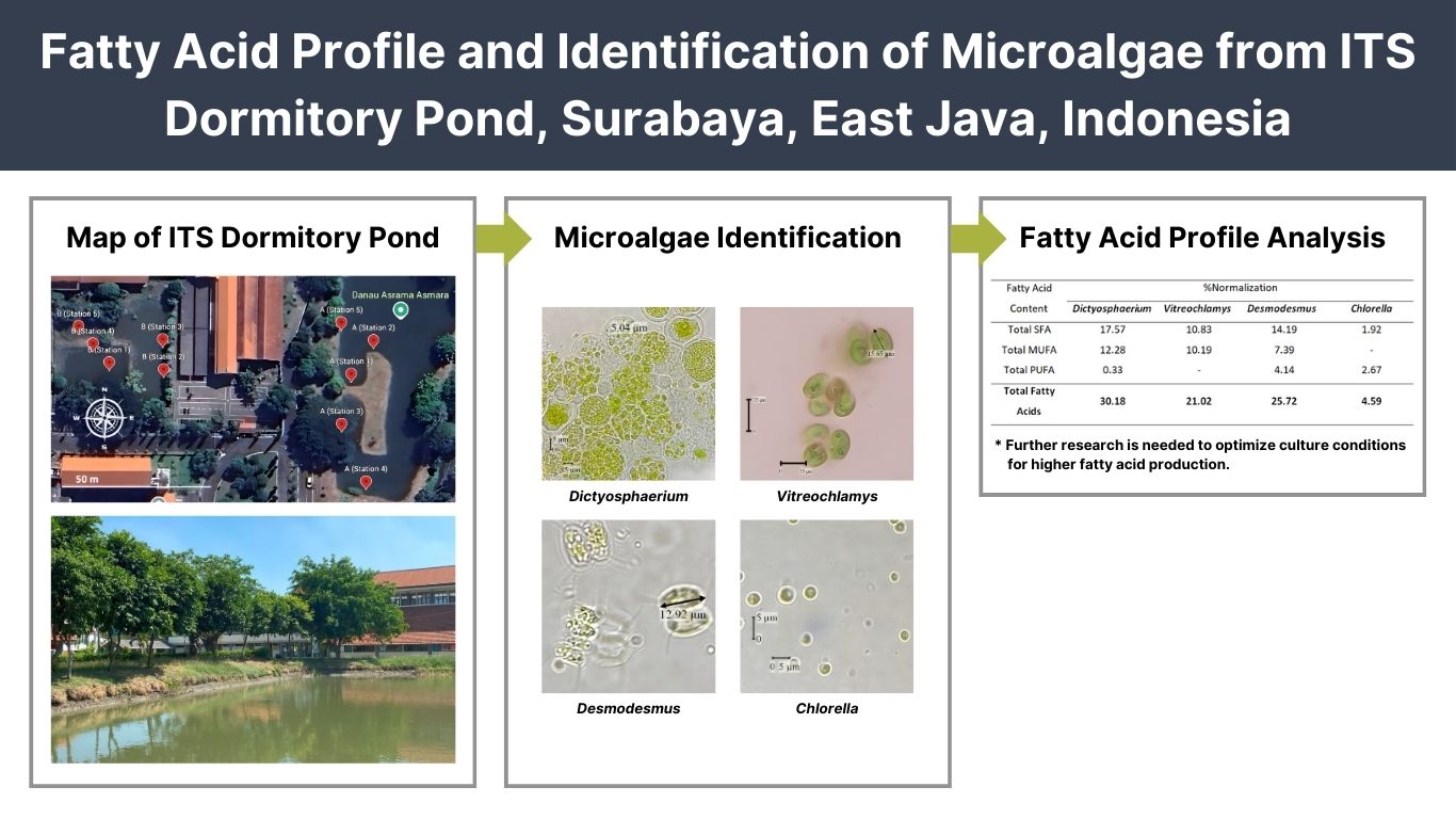
This research was conducted from June 2023 to July 2024. Water samples were taken from ITS Dormitory Pond (Figure 1; Table 1). Samples were observed in the Bioscience and Plant Technology Laboratory, Department of Biology, Faculty of Science and Data Analytics, ITS, Surabaya. Microalgae fatty acid profile test was carried out in the Faculty of Pharmacy, Airlangga University, Surabaya.
Procedures
Preparation of Agar Medium
Microalgae were isolated using Bold’s Basal Medium (BBM) agar medium following the protocol of (Ardiansyah et al., 2018). The medium was sterilized using an autoclave at 121 °C and 1.5 atm pressure for 30 minutes. The agar medium was poured into Petri dishes, sealed with plastic wrap, and allowed to solidify before use (Lee et al., 2014).
Microalgae Sampling, Isolation, and Cultivation
Microalgae were sampled using an active method from two ponds, with five sampling points per pond. Water was filtered through a 35 μm plankton net, and the samples were stored in labeled bottles and transported to the laboratory (Saputro et al., 2019).
Subsequently, the water samples were diluted via serial dilution (10-1 to 10-S) by transferring 1 mL of the sample sequentially into test tubes with 9 mL of distilled water (Chaidir et al., 2016). Purification of microalgae samples was conducted using the streak plate method. Petri dishes were incubated at 27 °C under 30 Watt fluorescent light for 7 - 14 days until microalgae grew. Isolates were further purified on agar to obtain homogeneous cultures. Pure cultures were transferred to a liquid medium in glass bottles for growth (Saputro et al., 2019).
Cultivation was conducted following the protocol of Kawaroe et al. (2010) by mixing 20% of microalgae culture with 80% of fresh liquid medium. Cultivation began at 20 mL and scaled to 5 L in media with pH 6, temperature 27 °C, light intensity 30 Watts, and continuous aeration (Kawaroe et al., 2010). Cell growth was monitored daily by measuring optical density at 680 nm (Lee et al., 2014). Harvesting occurred during the early
stationary phase, identified via the growth curve, to maximize metabolic compound accumulation, including lipids. Microalgae were separated from the medium using high-speed centrifugation (Chen et al., 2011); (Ferreira et al., 2019).
Figure 1.The location of microalgae sampling in ITS Dormitory Pond, Surabaya, Indonesia
No. |
Station | Coordinate |
||
|---|---|---|---|---|
1. |
Pond A (Station 1) |
7°17'17"S | 112°47'32"E |
|
2. |
Pond A (Station 2) |
7°17'17"S | 112°47'32"E |
|
3. |
Pond A (Station 3) |
7°17'18"S | 112°47'32"E |
|
4. |
Pond A (Station 4) |
7°17'19"S | 112°47'32"E |
|
5. |
Pond A (Station 5) |
7°17'17"S | 112°47'32"E |
|
6. |
Pond B (Station 1) |
7°17'17"S | 112°47'29"E |
|
7. |
Pond B (Station 2) |
7°17'17"S | 112°47'30"E |
|
8. |
Pond B (Station 3) |
7°17'17"S | 112°47'30"E |
|
9. |
Pond B (Station 4) |
7°17'17"S | 112°47'29"E |
|
10. |
Pond B (Station 5) |
7°17'17"S | 112°47'29"E |
|
Morphological Identification
Morphological identification of isolated microalgae was done microscopically (Chaidir et al., 2016). Samples were placed on a glass slide, added with distilled water, and observed under an Olympus BX71 microscope with 1,000x magnification and immersion oil. Identification was based on characteristics, such as cell color, shape, size, and the presence of flagella and pyrenoids, using the books titled “Freshwater Algae: Identification, Enumeration and Use as Bioindicators” (Bellinger & Sigee, 2015) and “Freshwater Algae of North America” (Wehr et al., 2015).
Microalgae Fatty Acid Profile Test
The fatty acid profile of microalgae was analyzed using GC-MS, following these steps: 1) fatty acid extraction, 2) fatty acid derivatization, and 3) GC- MS analysis (Chiu & Kuo, 2020). For extraction, 4 mL of hexane was added to 100 mg of the sample, vortexed for 2 minutes, and centrifuged at 3000 rpm for 4 minutes. The hexane extract was transferred to a derivatization tube, evaporated under nitrogen, and treated with 1 mL of 2% NaOH at 90 °C for 5 minutes. After cooling, derivatization was performed by adding 1 ml of BF3 and heating for 30 minutes. Extraction with 2 mL of hexane was followed by GC-MS analysis (Griffiths et al., 2010).
The GC-MS analysis was performed using an Agilent 7890B Network GC System equipped with an Agilent 5977B MSD detector. Separation was achieved using an Agilent HP-5MS column (0.25 mm × 30 m × 0.25 μm). The inlet temperature was set to 290 °C, and the oven temperature was programmed as follows: 170 °C (1 minute),
percentage of total identified fatty acids to provide a normalized profile for each microalgal isolate (Patel et al., 2018). The relative abundance of each fatty acid was determined using the peak area
\documentclass{article} \usepackage{amsmath} \begin{document} \displaystyle \% \text{ Normalization } = \left( \frac{\text{Peak Area of individual Fatty Acid}}{\text{Total Peak Area of All Fatty Acid}} \right) \times 100 \end{document}
normalization method, calculated as follows:
Data Analysis
The data obtained from the study were analyzed using an exploratory descriptive method to identify microalgae types from each sampling location. Morphological characteristics were examined under a light microscope, and identification was conducted using taxonomic keys. The fatty acid profile test was conducted on successfully cultivated microalgae cells using GC-MS. Peak identification was performed by comparing the mass spectra with reference spectra from the Wiley 8.0 library, and the relative abundance of each fatty acid was determined using peak area normalization. Data were displayed in tabular form and interpreted based on the sampling locations.
RESULTS AND DISCUSSION
Microalgae Isolates
Four distinct microalgae isolates were obtained from the ITS Dormitory Pond (Table 2). The isolation process involved serial dilution and the streak plate method, repeated until pure isolates were obtained, followed by periodic microscopic observations to confirm the purity of each isolate (Fernandez-Vanenzuela et al., 2021).
| No. | Isolate Code | Sampling Location | Color |
|---|---|---|---|
| 1. | HDB3a | Pond B (Station 3) | Green |
| 2. | 16HtDB2b | Pond B (Station 2) | Dark Green |
| 3. | 17HtDB2b | Pond B (Station 2) | Dark Green |
| 4. | KD3a | Pond B (Station 3) | Yellow |
Morphological Identification of Microalgae Isolates
The isolate HDB3a was identified as having round cells with diameters ranging from 4 μm to 10 μm. These cells formed small colonies of 4, 8, 16, or more cells, embedded in a transparent mucilaginous matrix. Each cell contained one or more cup-shaped chloroplasts with chlorophyll, giving them a green appearance. Pyrenoids were present within the chloroplasts, playing a role in carbon storage and metabolism (Figure 2, A1). The morphological characteristics of the isolate align with those described by (Bellinger & Sigee, 2015), particularly the spherical shape, mucilaginous colony formation, and cup-shaped chloroplasts, confirming its classification within the genus Dictyosphaerium. (Krienitz & Bock, 2010) also reported that Dictyosphaerium species typically form colonies as a defense mechanism against environmental stress. Additionally, (Bock et al., 2011) found that Dictyosphaerium-like species develop colonies through mucilaginous stalks originating from remnants of the mother cell, suggesting possible polyphyly within the Chlorellaceae family. (Mikhailyuk et al., 2020) further demonstrated the genus’s morphological diversity by identifying Dictyosphaerium-like forms in terrestrial habitats. The presence of this isolate in a pond affected by domestic waste suggested its ability to thrive in nutrient-rich conditions. Its colony formation and chloroplast structures further support its adaptability to environmental changes.
Based on observations, the isolate coded 16HtDB2b had oval-shaped cells with diameters ranging from 10 to 17 μm. The transparent and smooth cell walls lacked any distinct ornamentation or structural modifications. A large, cup-shaped chloroplast occupied most of the cell’s interior, containing chlorophyll pigments and pyrenoids, which functioned as storage centers for photosynthetic products, such as starch. These microalgae exhibited motility, suggesting the presence of flagella, although the flagella were not clearly observed (Figure 2, B1). The morphological characteristics of this isolate align with the description of the genus Vitreochlamys provided by Wehr et al. (2015). Vitreochlamys is a unicellular green flagellate with a smooth, swollen cell wall that separates from the protoplast and features a prominent anterior papilla. Vegetative cells are solitary, biflagellate, and broadly spindle-shaped, pyriform, or ellipsoidal. They typically measure 24 - 34 μm in length and 10 - 22 μm in width, with flagella that are equal to or slightly shorter than the cell length. Two contractile vacuoles are positioned near the base of the flagella. The chloroplast has multiple lobes and contains 4 - 8 similarly sized pyrenoids, which are ellipsoidal, ovoid, or bean- shaped. These pyrenoids are surrounded by two large starch grains and are scattered throughout the chloroplast.
Observations showed that the isolate coded 17HtDB2b had ellipsoid or cylindrical cells, measuring approximately 10 - 30 μm in length and 5 - 15 μm in width. The cell walls featured a complex structure with prominent spines or projections at both ends. The isolate formed colonies of 2 cells to 4 cells arranged in linear or flat formations. These microalgae were non-motile, as they lacked flagella (Figure 2, C1). The morphological characteristics of this isolate are consistent with the genus Desmodesmus, as described by (Bellinger & Sigee, 2015). The spines in Desmodesmus serve as a defense mechanism against microzooplankton predation, as their rigid structure makes ingestion difficult (Trainor, 1996). Additionally, the spines enhance buoyancy, helping the cells remain in the photic zone for optimal photosynthesis (Zhu et al., 2018). The surface morphology of Desmodesmus species, observed under SEM, varied significantly. For example, D. armatus exhibited a warty surface, D. quadricauda had a net-like structure, and D. serratus displayed a star-like pattern. Many Desmodesmus species show phenotypic plasticity, alternating between unicellular and colonial forms, as well as spiny and non-spiny variants. Environmental factors influence these transitions; for instance, D. subspicatus and D. armatus develop spiny colonies under low nutrient conditions, while unicellular forms dominate in phosphorus- or nitrogen-rich environments. When iron availability is low, both colonies and unicellular forms become non-spiny (Shubert et al., 2014).
The isolate coded KD3a consisted of small, round, single cells with diameters ranging from 2 μm to 10 μm. Each cell contained a large, parietal chloroplast occupying most of the cell volume, giving it a green color due to the presence of chlorophyll. The chloroplasts also contained pyrenoids, which facilitate photosynthesis. These microalgae were non-motile as they lacked flagella (Figure 2, D1). The morphological characteristics of this isolate align with those of the genus Chlorella, as described by (Bellinger & Sigee, 2015). Chlorella species are unicellular green algae characterized by spherical or ovoid cells, typically measuring 2 - 10 μm in diameter. Their robust cell walls are composed of cellulose and pectin, providing structural integrity (Takeda, 2004). The genus is distinguished by a single, cup-shaped chloroplast with a prominent pyrenoid, essential for carbon fixation and starch storage. Chlorella is commonly found in nutrient-rich aquatic environments, but can be easily overlooked due to its small size. Its adaptability to various environmental conditions, including polluted waters, makes it a significant genus in both ecological and biotechnological studies (Bellinger & Sigee, 2015).
Figure 2.Observed microalgae in this study that have been identified by comparing to literatures
Fatty Acid Profile Analysis
The harvesting of microalgal isolates was performed prior to conducting fatty acid profile analysis. The optimal harvest time was determined based on the growth curve of the microalgae, specifically during the early stationary phase. This phase is crucial as it marks the onset of metabolite accumulation, including lipids. During the early stationary phase, cell density reaches a constant and maximum level, resulting in the highest biomass concentration (Ferreira et al., 2019). The early stationary phase can be observed through the microalgae growth curve Figure 3. According to the growth curve, the harvest times for the microalgae were as follows: Dictyosphaerium was harvested on day 16, Vitreochlamys on day 15, and both Desmodesmus and Chlorella on day 13. The study results indicated that the microalgal species with the highest total fatty acid content was Dictyosphaerium (30.18%), followed by Desmodesmus (25.72%), Vitreochlamys (21.02%), and Chlorella (4.59%). The fatty acid composition of microalgae isolates from the ITS Dormitory Pond Waters showed significant variation. The fatty acid profile test results indicated that most isolates had high fatty acid content, particularly elaidic acid (C18:1), palmitic acid (C16:0), and stearic acid (C18:0) Table 3.
Figure 3.Chromatogram of fatty acid profile
| No. | Retention time | Component name | % Normalization | |||
|---|---|---|---|---|---|---|
| Dictyosphaerium | Vitreochlamys | Desmodesmus | Chlorella | |||
1. |
3.40 |
Lauric Acid (C12:0) |
0.11 |
- |
2.33 |
- |
2. |
6.04 |
Myristic Acid (C14:0) |
0.85 |
0.52 |
1.41 |
0.01 |
3. |
8.20 |
Myristic Acid (C14:0) |
0.20 |
- |
- |
- |
4. |
8.17 |
Pentadecanoic Acid (C5:0) |
- |
- |
0.26 |
- |
5. |
10.20 |
Hexadienoic Acid (C6:2)** |
- |
- |
0.32 |
0.22 |
6. |
10.57 |
Palmitic Acid (C16:0) |
- |
- |
1.02 |
- |
7. |
11.40 |
Palmitic Acid (C16:0) |
9.32 |
6.83 |
6.49 |
1.21 |
8. |
14.52 |
Margaric Acid (C17:0) |
- |
- |
0.16 |
- |
9. |
16.41 |
Linoleic Acid (C18:2)** |
0.33 |
- |
2.46 |
0.48 |
10. |
16.62 |
α-Linoleic Acid (C18:3(ω-3))** |
- |
- |
- |
1.97 |
11. |
16.70 |
Elaidic Acid (C18:1)* |
12.28 |
10.19 |
7.39 |
- |
12. |
17.27 |
Stearic Acid (C18:0) |
5.52 |
2.49 |
2.47 |
0.44 |
13. |
20.17 |
Arachidonic Acid (C20:4(ω-6))** |
- |
- |
0.44 |
- |
14. |
20.55 |
Eicosatrienoic Acid (C20:3(ω-3))** |
- |
- |
0.22 |
- |
15. |
21.59 |
Arachidic Acid (C20:0) |
0.54 |
0.35 |
0.05 |
0.26 |
16. |
23.87 |
Eicosapentaenoic Acid (C20:5(ω-3))** |
- |
- |
0.70 |
- |
17. |
25.20 |
Behenic Acid (C22:0) |
1.03 |
0.64 |
- |
- |
Total SFA |
17.57 |
10.83 |
14.19 |
1.92 |
||
Total MUFA |
12.28 |
10.19 |
7.39 |
- |
||
Total PUFA |
0.33 |
- |
4.14 |
2.67 |
||
Total Fatty Acids |
30.18 |
21.02 |
25.72 |
4.59 |
||
Dictyosphaerium exhibited the highest saturated fatty acid (SFA) content among the four microalgae species, with a total of 17.57% comprising lauric acid, myristic acid, palmitic acid, stearic acid, arachidic acid, and behenic acid. The species also showed the highest level of monounsaturated fatty acids (MUFA), at 12.28%, predominantly as elaidic acid. Conversely, Dictyosphaerium had a lower content of polyunsaturated fatty acids (PUFA), amounting to 0.33%, primarily as linoleic acid (Figure 4, A). In contrast, (Nayana et al., 2018) reported that Dictyosphaerium contained the highest levels of SFA, specifically palmitic acid (22.64%). The MUFA accounted for 44.24%, including oleic acid (38.08%) and palmitoleic acid (6.16%). The PUFA content was 8.93%, with linoleic acid being the predominant PUFA. The discrepancy between the results of this study and those reported by Nayana et al. (2018) may be attributed to differences in cultivation conditions, which influence the composition of fatty acids produced during growth.
Vitreochlamys exhibited an SFA content of 10.83%, consisting of myristic acid, palmitic acid, stearic acid, arachidic acid, and behenic acid. The MUFA content was 10.19%, predominantly as elaidic acid, with no detected PUFA (Figure 4, B). Research on the fatty acid profile of the genus Vitreochlamys is currently limited. Comparable studies within the family Chlamydomonadaceae can be observed, such as in Chlamydomonas sp., which contains SFA, including myristic acid (2.62%) and palmitic acid (78.35%). This species also has MUFA, like palmitoleic acid (4.26%) and oleic acid (2.48%), and PUFA, such as linoleic acid (3.49%) and arachidonic acid (3.18%). Palmitic acid (C16:0) is the most abundant fatty acid in Chlamydomonas sp., accounting for about 85% of the total fatty acid content, with 9% being PUFA (Morando-Grijalva et al., 2020).
Desmodesmus exhibited an SFA content of 14.19%, which includes lauric acid, myristic acid, pentadecanoic acid, palmitic acid, margaric acid, stearic acid, and arachidic acid. The MUFA content was 7.39%, primarily as elaidic acid. Notably, Desmodesmus had the highest PUFA content among the four species analyzed, with a total of 4.14%, including hexadecanoic acid, linoleic acid, arachidonic acid, eicosatrienoic acid, and eicosapentaenoic acid (Figure 4, C). In other studies, Desmodesmus was found to contain different compounds under autotrophic (Bourdeau et al., 2017), heterotrophic (Deschênes et al., 2015), and mixotrophic growth conditions (Okpozu et al., 2019).
Chlorella exhibited an SFA content of 1.92%, which included myristic acid, palmitic acid, stearic acid, and arachidic acid. The PUFA content was 2.67%, comprising hexadecanoic acid, linoleic acid, and α-linolenic acid. Notably, no MUFA were detected in Chlorella in this research (Figure 4, D). According to (Rushan et al., 2021), Chlorella cultivated in BG-11, JM, and BBM media displayed typical fatty acid compositions found in microalgal biomass, including palmitic acid (C16:0), stearic acid (C18:0), oleic acid (C18:1), linoleic acid (C18:2), and linolenic acid (C18:3). The highest SFA content was observed in Chlorella cultivated with BBM, followed by JM and BG-11 media. The highest SFA composition in BBM-cultivated Chlorella was attributed to C16:0 (40.74%) and C18:0 (33.33%) of total fatty acid methyl esters (FAME). The highest UFA composition was found in BBM due to the presence of C18:1 (11.11%), C18:2 (7.41%), and C18:3, (3.7%) of total FAME. This shows that using different media will produce different fatty acid contents (Rushan et al., 2021).
Figure 4.Chromatogram of fatty acid profile
CONCLUSION
This study successfully identified various microalgae from the waters of ITS Dormitory Pond in Surabaya, including Dictyosphaerium, Vitreochlamys, Desmodesmus, and Chlorella. The fatty acid profiles of the isolated microalgae species varied. Dictyosphaerium was found to have a saturated fatty acid (SFA) content of 17.57%, monounsaturated fatty acid (MUFA) content of 12.28%, and polyunsaturated fatty acid (PUFA) content of 0.33%. Vitreochlamys exhibited an SFA content of 10.83% and a MUFA content of 10.19%, with no detected PUFA. Desmodesmus showed an SFA content of 14.19%, MUFA content of 7.39%, and PUFA content of 4.14%. Chlorella had an SFA content of 1.92% and a PUFA content of 2.67%, with no detected MUFA. Future research should focus on optimizing culture conditions to enhance lipid accumulation and improve fatty acid composition for commercial applications. Investigating genetic and metabolic pathways could further increase lipid productivity and tailor fatty acid profiles for specific industrial uses. Additionally, evaluating large-scale cultivation feasibility and sustainability aspects will be essential for developing microalgae-based bioresources in biofuels, nutraceuticals, and pharmaceuticals.
References
- Abdelkarim O.H., Wijffels R.H., Barbosa M.J.. Microalgal lipid production: A comparative analysis of Nannochloropsis and Microchloropsis strains. J Appl Phycol. 2024; 37(1):15-34.
- Ardiansyah Orlando, AM Rahman, A Prihantini, NB Nasruddin. Effect of aeration in simple photobioreactor system for biomass production of Synechococcus sp. 2018.
- Bellinger E.G., Sigee D.C.. John Wiley & Sons: New Delhi (IN; 2015.
- Bock C., Pröschold T., Krienitz L.. Updating the genus Dictyosphaerium and description of Mucidosphaerium gen. nov. Trebouxiophyceae) based on morphological and molecular data. J Phycol. 2011; 47(3):638-52.
- Bourdeau N., Bélanger-Lépine F., Adjallé K., Dubois-Caléro N., Dosnon-Olette R., Samson G., Barnabé S.. Mixotrophic cultivation of an algae-bacteria consortium in aluminium smelter wastewaters. Ind Biotech. 2017; 13(5):260-9.
- Chaidir Z., Fadjria N., Armaini Zainul, R.. Isolation and molecular identification of freshwater microalgae in Maninjau Lake West Sumatra. Sch Res Lib. 2016; 8(20):177-87.
- Chen W., Li T., Du S., Chen H., Wang Q.. Microalgal polyunsaturated fatty acids: Hotspots and production techniques. Front Bioeng Biotechnol. 2023; 11(1146881):1-22.
- Chen Y.C., Yeh K.L., Aisyah R., Lee D.J., Chang J.S.. Cultivation, photobioreactor design and harvesting of microalgae for biodiesel production: A critical review. Bioresour Technol. 2011; 102:71-81.
- Chiu H., Kuo C.. Gas chromatography-mass spectrometry-based analytical strategies for fatty acid analysis in biological samples. J Food Drug Anal. 2020; 28(1):60-73.
- Deschênes J., Boudreau A., Tremblay R.. Mixotrophic production of microalgae in pilot-scale photobioreactors: Practicability and process considerations. Algal Res. 2015; 10:80-6.
- Microalgae: A promising source of valuable bioproducts. Biomolecules. 2020; 10(8)
- Elisabeth B., Rayen F., Behnam T.. Microalgae culture quality indicators: A review. Crit Rev Biotechnol. 2021; 41(4):451-73.
- Fernandez-Vanenzuela S., Ruvalcaba F.C., Beltran-Rocha J.C., Claudio P.M.S.. Isolation and culturing axenic microalgae: Mini–Review. Open Microbiol J. 2021; 15(1):111-19.
- Ferreira G.F., Pinto L.F.R., Carvalho P.O., Coelho M.B., Eberlin M.N., Filho R.M., Fregolente L.V.. Biomass and lipid characterization of microalgae genera Botryococcus, Chlorella, and Desmodesmus aiming high-value fatty acid production. Biomass Convers Biorefin. 2019; 11:1675-89.
- Griffiths M.J., Hille R.P., Harrison S.T.L.. Selection of direct transesterification as the preferred method for assay of fatty acid content of microalgae. Lipids. 2010; 45:1053-60.
- Kawaroe M., Prartono T., Rachmat A., Sari D.W., Augustine D.. IPB Press: Bogor (ID; 2010.
- Krienitz L., Bock C.. Polyphiletic origin of the Dictyosphaerium Morphotype within Chlorellaceae (Trebouxiophyceae. J Phycol. 2010; 46(3):559-63.
- Lee K., Eisterhold M.L., Rindi F., Palanisami S., Nam P.K.. Isolation and screening of microalgae from natural habitats in the midwestern United States of America for biomass and biodiesel sources. J Nat Sci Biol Med. 2014; 5(2):333-39.
- Mazidah R., Mulyadi A., Nasution S.. Pusat Penelitian Lingkungan Hidup: Riau (ID; 2013.
- Mikhailyuk T., Holzinger A., Tsarenko P., Glaser K.. Dictyosphaerium‐like morphotype in terrestrial algae: What is Xerochlorella (Trebouxiophyceae, Chlorophyta)?. J Phycol. 2020; 56(3):1-16.
- Milano J., Ong H.W., Masjuki H.H., Chong W.T., Lam M.K., Loh P.K., Vellayan V.. Microalgae biofuels as an alternative to fossil fuel for power generation. Renew Sustain Energy Rev. 2016; 58:180-97.
- Morando-Grijalva C.A., Vázquez-Larios A.L., Alcántara-Hernández R.J., Ortega-Clemente L.A., Robledo-Narváez P.N.. Isolation of a freshwater microalgae and its application for the treatment of wastewater and obtaining fatty acids from tilapia cultivation. Environ Sci Pollut Res. 2020; 27(23):28575-584.
- Nayana B., Venkatesh H.N., Sudharsana T.N., Manjunath K., Mohana D.C.. Isolation and utilization of Dictyosphaerium ehrenbergianum Nageli for biodiesel production: Lipid extraction and biodiesel property analysis. Biofuels. 2018; 11:885-92.
- Okpozu O.O., Ogbonna I.O., Ikwebe J., Ogbonna J.C.. Phycoremediation of cassava wastewater by Desmodesmus armatus and the concomitant accumulation of lipids for biodiesel production. Bioresour Technol Rep. 2019; 7(100255):1-7.
- Patel M.K., Das S., Thakur J.K.. GC-MS-based analysis of methanol: Chloroform-extracted fatty acids from plant tissues. Bio Protoc. 2018; 8(18):1-9.
- Rushan N.H., Mat Yasin N.H., Said F.M.. The effect of culture medium on the oil yield and fatty acid methyl ester of freshwater microalgae Chlorella vulgaris. Chem Eng Comms. 2021; 208(4):592-600.
- Saputro T.B., Purwani K.I., Ermavitalini D., Saifulloh A.F.. Isolation of high lipid content microalgae from Wonorejo River, Surabaya, Indonesia and its identification using rbcL marker gene. Biodiversitas. 2019; 20(5):1380-88.
- Shang X., Yang Y., Zan Y., Sun Z., Lu Z., Sun J.. Front Mar Sci. 2024:1-9.
- Shubert E., Wilk-Wozniak E., Ligeza S.. An autecological investigation of Desmodesmus: implications for ecology and taxonomy. Plant Ecol Evol. 2014; 147(2):202-12.
- Takeda H.. Sugar composion of cell wall and the taxonomy of Chlorella (Chlorophyceae. J Phycol. 2004; 27(2):224-32.
- Trainor F.R.. Reproduction in Scenedesmus. Algae. 1996; 11(2):183-201.
- Wehr J.D., Sheath R.G., Kociolek J.P.. Academic Press: San Diego (US; 2015.
- Wu J., Gu X., Yang D., Xu S., Wang S., Chen X., Wang Z.. Bioactive substances and potentiality of marine microalgae. Food Sci Nutr. 2021; 9(9):5279-92.
- Zhu W., Li Y., Fei X., Deng X.. Nutrient removal and algal community variation from urban river with the isolated microalgal strains Chlorella sp. and Scenedesmus sp. J Water Resour Prot. 2018; 10(9):884-95.
Ardiansyah SR, Orlando AM, Rahman A, Prihantini NB, Nasruddin. 2018. Effect of aeration in simple photobioreactor system for biomass production of synechococcus sp. (cyanobacteria) HS-7 and HS-9 as biofuel feedstock. E3S Web Conf 67(02006):1-6. DOI: https://doi.org/10.1051/e3sconf/20186702006
Armelia A, Djarot IN, Paminto AK, Nurfaiz I, Nuha, Handayani T. 2023. Analisis Limbah Media Zarrouk Modifikasi yang Digunakan untuk Budidaya Spirulina platensis dan Analisis Kualitas Biomassanya sebagai Bahan Pangan Fungsional [Analysis of Modified Zarrouk Media Waste Used for Cultivating Spirulina platensis and Analysis of Biomass Quality as a Functional Food Ingredient]. J Teknol Lingkungan 24(2):315-322. DOI: https://doi.org/10.55981/jtl.2023.300
Bellinger EG, Sigee DC. 2015. Freshwater Algae: Identification, Enumeration and Use as Bioindicators. New Delhi: John Wiley & Sons, Ltd. DOI: https://doi.org/10.1002/9781118917152
Bourdeau N, Bélanger-Lépine F, Adjallé K, Dubois-Caléro N, Dosnon-Olette R, Samson G, Barnabé S. 2017. Mixotrophic cultivation of an algae-bacteria consortium in aluminium smelter wastewaters (Quebec, Canada): high nitrogen concentration increases overall lipid production. Ind Biotech 13(5):260-269. DOI: https://doi.org/10.1089/ind.2017.0006
Chaidir Z, Fadjria N, Armaini, Zainul R. 2016. Isolation and Molecular Identification of Freshwater Microalgae in Maninjau Lake West Sumatra. Sch Res Lib 8(20):177-187.
Chen W, Li T, Du S, Chen H, Wang Q. 2023. Microalgal polyunsaturated fatty acids: Hotspots and production techniques. Front Bioeng Biotechnol 11(1146881), pp.1-22. DOI: https://doi.org/10.3389/fbioe.2023.1146881
Chen YC, Yeh KL, Aisyah R, Lee DJ, Chang JS. 2011. Cultivation, photobioreactor design and harvesting of microalgae for biodiesel production: A critical review. Bioresour Technol 102:71-81. DOI: https://doi.org/10.1016/j.biortech.2010.06.159
Chiu H, Kuo C. 2020. Gas chromatography-mass spectrometry-based analytical strategies for fatty acid analysis in biological samples. J Food Drug Anal 28(1):60-73. DOI: https://doi.org/10.1016/j.jfda.2019.10.003
Deschênes J, Boudreau A, Tremblay R. 2015. Mixotrophic production of microalgae in pilot-scale photobioreactors: practicability and process considerations. Algal Res 10:80-86. DOI: https://doi.org/10.1016/j.algal.2015.04.015
Dolganyuk V, Belova D, Babich O, Prosekov A, Ivanova S, Katserov D, ..., Sukhikh S. 2020. Microalgae: A Promising Source of Valuable Bioproducts. Biomolecules 10(8):1153. DOI: https://doi.org/10.3390/biom10081153
Elisabeth B, Rayen F, Behnam T, 2021. Microalgae culture quality indicators: a review. Crit Rev Biotechnol 41(4):451-473. DOI: https://doi.org/10.1080/07388551.2020.1854672
Ermavitalini D, Dwirejeki S, Nurhatika S, Saputro TB. 2019. Pengaruh Kombinasi Cekaman Nitrogen dan Fotoperiode terhadap Biomassa, Kandungan Kualitatif Triasilgliserol dan Profil Asam Lemak Mikroalga Nannochloropsis sp. [Effect of Combination of Nitrogen Stress and Photoperiod on Biomass, Qualitative Triacylglycerol Content and Fatty Acid Profile of the Microalga Nannochloropsis sp.]. Indo Chim Acta 4(1):32-49.
Ferreira GF, Pinto LFR, Carvalho PO, Coelho MB, Eberlin MN, Filho RM, Fregolente LV. 2019. Biomass and lipid characterization of microalgae genera Botryococcus, Chlorella, and Desmodesmus aiming high-value fatty acid production. Biomass Convers Biorefin 11:1675–1689. DOI: https://doi.org/10.1007/s13399-019-00566-3
Gildantia A, Ferniah RS, Budiharjo A, Suprihadi A, Zainuri M, Kusumaningrum HP. 2022. Identifikasi Spesies Mikroalga dari BBPBAP Jepara secara Morfologi dan Molekuler menggunakan 18S rDNA [Identification of Microalgae Species from BBPBAP Jepara Morphologically and Molecularly using 18S rDNA]. BULOMA 11(2):167-176. DOI: https://doi.org/10.14710/buloma.v11i2.39703
Handra I, Syafrizayanti, Chaidir Z. 2019. Isolasi dan Identifikasi Mikroalga sebagai Sumber Antioksidan dari Perairan Tirtasari Sonsang, Agam, Sumatera Barat [Isolation and Identification of Microalgae as a Source of Antioxidants from the Waters of Tirtasari Sonsang, Agam, West Sumatra]. J Chimica et Natura Acta 7(1):7-13. DOI: https://doi.org/10.24198/cna.v7.n1.20076
Hernandi R, Dharma A, Armaini A., 2019. Penapisan, isolasi, dan karakterisasi mikroalga yang berpotensi sebagai sumber biodiesel dari perairan Danau Kerinci, Jambi [Screening, isolation and characterization of microalgae that have the potential as a source of biodiesel from the waters of Lake Kerinci, Jambi]. J Litbang Ind 9(1):41-49. DOI: https://doi.org/10.24960/jli.v9i1.4326.41-49
Hindarti F, Ayuningtyas E. 2020. Pengembangan Teknik Kultivasi Spirulina sp. sebagai Sumber Biomassa Energi Terbarukan dalam Fotobioreaktor Airlift [Development of Spirulina sp Cultivation Techniques. as a Renewable Biomass Energy Source in Airlift Photobioreactors]. J Enerlink 16(1):17-24. DOI: https://doi.org/10.29122/jel.v16i1.4578
Kawaroe M, Prartono T, Rachmat A, Sari DW, Augustine D. 2010. Mikroalga : Potensi dan Pemanfaatannya Untuk Produksi Bio Bahan Bakar [Microalgae: Potential and Use for Biofuel Production]. Bogor: IPB Press.
Krienitz L, Bock C. 2010. Polyphiletic Origin of the Dictyosphaerium Morphotype within Chlorellaceae (Trebouxiophyceae). J Phycol 46(3):559-563. DOI: https://doi.org/10.1111/j.1529-8817.2010.00813.x
Lee K, Eisterhold ML, Rindi F, Palanisami S, Nam PK. 2014. Isolation and screening of microalgae from natural habitats in the midwestern United States of America for biomass and biodiesel sources. J Nat Sc Biol Med 5(2):333-339. DOI: https://doi.org/10.4103/0976-9668.136178
Mazidah R, Mulyadi A, Nasution S. 2013. Tingkat Pencemaran Perairan Danau Buatan Pekanbaru Ditinjau dari Parameter Fisika, Kimia, dan Biologi [Pekanbaru Artificial Lake Water Pollution Level Seen from Physical, Chemical and Biological Parameters]. Riau: Pusat Penelitian Lingkungan Hidup.
Morando-Grijalva CA, Vázquez-Larios AL, Alcántara-Hernández RJ, Ortega-Clemente LA, Robledo-Narváez PN. 2020. Isolation of a freshwater microalgae and its application for the treatment of wastewater and obtaining fatty acids from tilapia cultivation. Environ Sci Pollut Res 27(23):28575-28584. DOI: https://doi.org/10.1007/s11356-020-08308-z
Nagappan S, Das P, AbdulQuadir M, Thaher M, Khan S, Mahata C, ..., Kumar G. 2021. Potential of microalgae as a sustainable feed ingredient for aquaculture. J Biotechnol 341:1-20. DOI: https://doi.org/10.1016/j.jbiotec.2021.09.003
Nayana B, Venkatesh HN, Sudharsana TN, Manjunath K, Mohana DC. 2018. Isolation and utilization of Dictyosphaerium ehrenbergianum Nageli for biodiesel production: lipid extraction and biodiesel property analysis. Biofuels 11:885-892. DOI: https://doi.org/10.1080/17597269.2018.1432270
Neinggolan AA, Eddiwan, Windarti. 2022. Identifikasi dan Isolasi Mikroalga dari Perairan Rawa Gambut di Kelurahan Air Hitam Kota Pekanbaru Provinsi Riau [Identification and Isolation of Microalgae from Peat Swamp Waters in Air Hitam Village, Pekanbaru City, Riau Province]. JSLA 3(2):1-10.
Okpozu OO, Ogbonna IO, Ikwebe J, Ogbonna JC. 2019. Phycoremediation of cassava wastewater by Desmodesmus armatus and the concomitant accumulation of lipids for biodiesel production. Bioresour Technol Rep 7(100255):1-7. DOI: https://doi.org/10.1016/j.biteb.2019.100255
Putri DS, Marianah, Ihromi S. 2018. Isolasi Mikroalga Laut dari Pantai Mapak Pulau Lombok [Isolation of Marine Microalgae from Mapak Beach, Lombok Island]. J Agrotek 5(2):91-95. DOI: https://doi.org/10.31764/agrotek.v5i2.699
Rozana E, Anwar SH, Sulaiman MI. 2021. Potensi Minyak Mikroalga dan Khamir sebagai Sumber Asam Lemak Esensial [Potential of Microalgae and Yeast Oils as Sources of Essential Fatty Acids]. Agrointek 31(3):332-342. DOI: https://doi.org/10.24961/j.tek.ind.pert.2021.31.3.332
Rushan NH, Mat Yasin NH, Said FM. 2021. The effect of culture medium on the oil yield and fatty acid methyl ester of freshwater microalgae Chlorella vulgaris. Chem Eng Comms 208(4):592-600. DOI: https://doi.org/10.1080/00986445.2020.1864622
Salindeho RSE, Budijono, Hendrizal A. 2022. Identifikasi dan Kelimpahan Mikroalga Dari Sungai Rawa Kawasan Taman Nasional Zamrud Kabupaten Siak [Identification and Abundance of Microalgae from the Rawa River in the Emerald National Park Area, Siak Regency]. JSLA 3(1):1-7.
Saputro TB, Purwani KI, Ermavitalini D, Saifulloh AF. 2019. Isolation of high lipid content microalgae from Wonorejo river, Surabaya, Indonesia and its identification using rbcL marker gene. Biodiversitas 20(5):1380-1388. DOI: https://doi.org/10.13057/biodiv/d200530
Sari AN, Hutabarat S, Soedarsono P. 2014. Struktur Komunitas Plankton pada Padang Lamun di Pantai Pulau Panjang, Jepara [Plankton Community Structure in Seagrass Fields on Panjang Island Beach, Jepara]. Diponegoro J Maquares 3(2):82-91.
Shang X, Yang Y, Zan Y, Sun Z, Lu Z, Sun J. 2024. Effects of temperature on the growth, total lipid content and fatty acid composition of Skeletonema dohrnii. Front Mar Sci 1-9. DOI: https://doi.org/10.3389/fmars.2024.1361157
Shubert E, Wilk-Wozniak E, Ligeza S. 2014. An autecological investigation of Desmodesmus: implications for ecology and taxonomy. Plant Ecol Evol 147(2):202-212. DOI: https://doi.org/10.5091/plecevo.2014.902
Trainor FR. 1996. Reproduction in Scenedesmus. Algae 11(2):183-201.
Wehr JD, Sheath RG, Kociolek JP. 2015. Freshwater Algae of North America. USA: Academic Press. DOI: https://doi.org/10.1016/B978-0-12-385876-4.00005-0
Zhu W, Li Y, Fei X, Deng X. 2018. Nutrient Removal and Algal Community Variation from Urban River with the Isolated Microalgal Strains Chlorella sp. and Scenedesmus sp. J Water Resour Prot 10(9):884-895. DOI: https://doi.org/10.4236/jwarp.2018.109051
Copyright (c) 2025 Alfiyyana Nurrahma Mawardani, Dini Ermavitalini, Triono Bagus Saputro, Farid Kamal Muzaki, Adi Setyo Purnomo, Hery Purnobasuki

This work is licensed under a Creative Commons Attribution-NonCommercial-NoDerivatives 4.0 International License.
Authors who publish with this journal agree with the following terms:
- Authors retain copyright and grant the journal right of first publication, with the work 1 year after publication simultaneously licensed under a Creative Commons attribution-noncommerical-noderivates 4.0 International License that allows others to share, copy and redistribute the work in any medium or format, but only where the use is for non-commercial purposes and an acknowledgement of the work's authorship and initial publication in this journal is mentioned.
- Authors are able to enter into separate, additional contractual arrangements for the non-exclusive distribution of the journal's published version of the work (e.g., post it to an institutional repository or publish it in a book), with an acknowledgement of its initial publication in this journal.
- Authors are permitted and encouraged to post their work online (e.g., in institutional repositories or on their website) prior to and during the submission process, as it can lead to productive exchanges, as well as earlier and greater citation of published work (See The Effect of Open Access).